
TAVR-vs-SAVR-1200x778.png

September 28, 2026 AbhijitHearty Life

Aortic valve disease can affect how efficiently the heart pumps blood throughout the body. One of the most common valve conditions is aortic stenosis, in which the aortic valve becomes narrowed and restricts blood flow from the heart. When the condition becomes severe, valve replacement may be recommended.

Two important treatment approaches are TAVI vs. SAVR. TAVI, also known as Transcatheter Aortic Valve Implantation, is a catheter-based procedure, while SAVR, or Surgical Aortic Valve Replacement, involves replacing the valve through open-heart surgery.

Understanding the differences between these procedures can help patients discuss appropriate treatment options with their cardiologist.

What Is Aortic Stenosis?

Aortic stenosis occurs when the aortic valve becomes narrowed, often because of calcium buildup, congenital abnormalities, or age-related valve degeneration. The narrowing makes it harder for the heart to push blood into the aorta.

Symptoms may include:

  • Breathlessness, particularly during physical activity
  • Chest discomfort or pain
  • Dizziness or fainting
  • Fatigue and reduced exercise capacity
  • Swelling in the legs or ankles
  • Palpitations in some patients

Severe or symptomatic aortic stenosis may require intervention. The appropriate aortic stenosis treatment depends on several factors, including valve anatomy, age, symptoms, overall health, surgical risk, and other medical conditions.

What Is TAVI?

TAVI stands for Transcatheter Aortic Valve Implantation. It is also commonly called TAVR, or Transcatheter Aortic Valve Replacement.

Instead of surgically opening the chest to remove the diseased valve, a replacement valve is delivered through a catheter. In many cases, the catheter is introduced through an artery in the groin and guided toward the heart.

The new valve is positioned inside the existing aortic valve and expanded into place. The replacement valve then takes over the function of the diseased valve.

Patients searching for a TAVR doctor in Hyderabad, TAVR expert in Hyderabad, or TAVI specialist in Hyderabad should discuss their valve anatomy and overall medical condition with an experienced structural heart team.

What Is SAVR?

SAVR means Surgical Aortic Valve Replacement. It is a conventional surgical procedure in which the damaged aortic valve is removed and replaced with a prosthetic valve.

The operation generally requires surgical access to the heart, and cardiopulmonary bypass is used during the procedure. Depending on the patient’s circumstances, either a mechanical or tissue valve may be selected.

Surgical aortic valve replacement remains an important treatment option for patients with severe aortic valve disease, particularly when surgery is considered more appropriate because of anatomical, clinical, or other factors.

TAVI vs. SAVR: Key Differences

The main difference between TAVI and SAVR is how the replacement valve is delivered and implanted.

Factor TAVI/TAVR SAVR
Procedure Catheter-based Surgical
Chest opening Usually no open-chest surgery Requires surgical access
Valve placement Replacement valve placed through a catheter Diseased valve surgically replaced
Recovery Often shorter Generally longer
Hospital stay Usually shorter, depending on recovery Usually longer
Anesthesia Depends on the individual procedure and centre Generally requires general anesthesia
Patient suitability Depends on anatomy, age, risk and clinical factors Depends on surgical risk, anatomy and other factors

These are general differences. The actual treatment plan should be based on an individual assessment rather than the procedure alone.

Benefits of TAVI

TAVI has changed the treatment approach for many patients with aortic stenosis. Some potential benefits include:

Less invasive treatment

TAVI does not usually require the same type of open-chest surgery associated with conventional valve replacement.

Shorter recovery for many patients

Because TAVI is less invasive, many patients can resume normal activities sooner than after major cardiac surgery, although recovery varies from person to person.

Option for patients with higher surgical risk

TAVI may be considered for certain patients for whom conventional surgery carries significant risk. However, suitability must be determined by a specialist heart team.

Catheter-based valve replacement

The procedure allows a replacement valve to be delivered directly to the heart through a catheter, avoiding surgical removal of the native valve in the traditional manner.

Benefits of SAVR

SAVR also has an established role in the treatment of aortic valve disease.

Established surgical approach

SAVR has been performed for many years and remains an important option for appropriate patients.

Treatment of complex anatomy

Certain anatomical conditions may make surgical valve replacement more suitable than a transcatheter procedure.

Opportunity to address other cardiac problems

If a patient requires another cardiac surgical procedure at the same time, surgery may provide an opportunity to address multiple problems during one operation.

Mechanical or tissue valve options

Depending on age, medical history, lifestyle and other factors, patients may be considered for mechanical or biological valve choices.

How Is the Right Treatment Chosen?

Choosing between TAVI and SAVR is not simply a matter of selecting the less invasive procedure. A detailed evaluation is necessary.

A heart team may consider:

  • Patient age and life expectancy
  • Severity of aortic stenosis
  • Symptoms and functional status
  • Aortic valve anatomy
  • Size and structure of the aortic root
  • Coronary artery anatomy
  • Surgical risk
  • Previous cardiac procedures
  • Kidney and lung function
  • Other medical conditions
  • Long-term valve considerations
  • Patient preferences

Imaging, particularly echocardiography and CT evaluation, can provide important information before the treatment decision is made.

What About Congenital Aortic Stenosis?

Congenital aortic stenosis can occur when a person is born with an abnormal aortic valve or related structural abnormality. The treatment approach depends on the patient’s age, valve anatomy, severity of obstruction and associated cardiac conditions.

Not every patient with congenital aortic stenosis requires valve replacement. Some may be managed with monitoring or other interventions, while severe disease may require a procedure.

This is why assessment by experienced aortic stenosis specialists is particularly important in patients with complex or congenital valve disease.

Recovery After TAVI and SAVR

Recovery varies considerably between patients.

After TAVI, patients are usually monitored for heart rhythm, valve function, vascular complications and other potential issues. Activity is gradually increased according to medical advice.

SAVR generally involves a longer recovery because it is major cardiac surgery. Patients may need more time for the surgical wound and chest to heal and may require a structured cardiac rehabilitation programme.

Regardless of the procedure, regular follow-up is important to monitor the function of the replacement valve and overall cardiovascular health.

Why Experience Matters in TAVI Treatment

TAVI requires detailed assessment of valve anatomy, vascular access, coronary anatomy and other structural features. Careful planning helps the heart team determine whether a transcatheter approach is appropriate.

Patients searching for a TAVR specialist in India, best TAVR doctors, or aortic stenosis specialists in Hyderabad should consider the doctor’s experience with structural heart procedures as well as the expertise of the multidisciplinary team and facilities available.

For patients seeking a best cardiologist in india, the treatment decision should be based on an individualized evaluation rather than a single procedure being considered suitable for everyone.

TAVI vs. SAVR: Which Treatment Is Suitable?

There is no single treatment that is appropriate for every patient with aortic stenosis.

TAVI vs. SAVR decisions require consideration of clinical history, valve anatomy, surgical risk, age, expected longevity, associated heart conditions and the patient’s treatment preferences.

A multidisciplinary heart team can assess these factors and explain the potential benefits and limitations of each approach. In some patients, TAVI may be appropriate, while in others, SAVR may provide the more suitable treatment strategy.

Early evaluation is important when symptoms such as breathlessness, chest discomfort, dizziness or reduced exercise tolerance develop. Timely assessment can help determine whether monitoring, medication or valve intervention is required.

Conclusion

Both TAVI and SAVR are established approaches for treating severe aortic valve disease. TAVI provides a catheter-based alternative to conventional surgery for appropriately selected patients, while SAVR continues to be an important option, particularly when surgical treatment is clinically appropriate.

The choice between TAVI vs. SAVR should be made after a detailed evaluation of the patient’s condition, valve anatomy, overall health and long-term treatment needs.

For evaluation and personalized guidance regarding TAVI, SAVR, and aortic stenosis treatment, consult Dr. C. Raghu, experienced interventional cardiologist in Hyderabad.


recovery-after-angioplasty-1200x777.png

September 21, 2026 AbhijitHealthcare

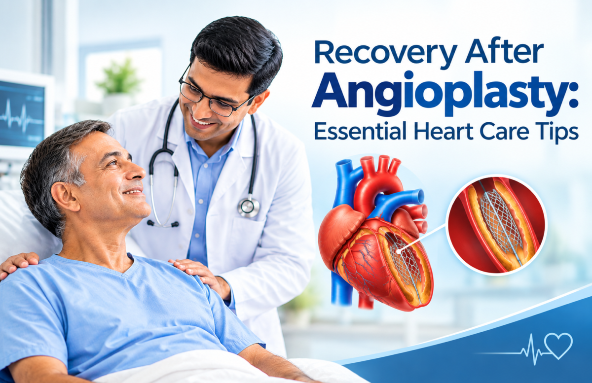
After a coronary angioplasty or PTCA stent procedure, the first concern for many patients is simple: What should be done now to protect the heart and avoid complications? Proper recovery after angioplasty involves more than allowing the catheter site to heal. Medicines, activity, diet, follow-up care, and management of coronary artery disease all influence long-term heart health.

This guide explains practical precautions after angioplasty, warning signs that need medical attention, and lifestyle measures patients and families can follow with guidance from the treating cardiologist.

Recovery After Angioplasty: What to Expect in the First Few Days
recovery after Angioplasty

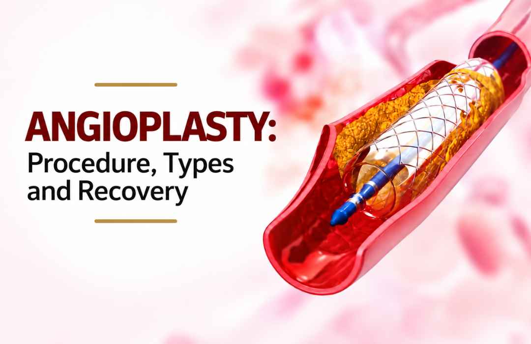
Coronary angioplasty, also called percutaneous coronary intervention or PCI, opens a narrowed or blocked coronary artery. A small balloon is used to widen the affected area, and a coronary stent is commonly placed to help keep the artery open.

Recovery varies depending on the reason for angioplasty, the patient’s overall health, the access site used, and whether the procedure was routine or complex. Some patients may return home the same day, while others require overnight observation.

Mild bruising or tenderness around the wrist or groin access site can occur. Patients should follow their discharge instructions closely instead of assuming that feeling better means the artery disease has been completely resolved.

PTCA Stent Medicines Must Be Taken as Prescribed

Medication adherence is one of the most important parts of recovery after a PTCA Stent procedure.

After stent placement, the cardiologist may prescribe antiplatelet medicines to reduce the likelihood of a clot forming inside the stent. The specific medicine and duration depend on the patient’s condition and treatment plan. Patients should never stop these medicines or change their dose without speaking with the treating cardiologist, even when they feel well.

Other prescribed medicines may help manage cholesterol, blood pressure, diabetes, angina, or other cardiovascular risk factors. Keeping an updated medication list can make follow-up appointments easier and reduce confusion for patients and family members.

Coronary Angioplasty Access-Site Care and Activity

The wrist or groin used for catheter insertion needs time to heal. Patients should keep the area clean and follow the hospital team’s instructions regarding dressing removal, bathing, and activity.

During early recovery:

  • Avoid heavy lifting and strenuous exercise until medically cleared.
  • Increase walking and routine activity gradually.
  • Watch the catheter site for increasing swelling, persistent bleeding, redness, or worsening pain.
  • Ask the cardiologist when driving, work, gym activity, and sexual activity can safely resume.
  • Attend cardiac rehabilitation if it is recommended.

A person’s timetable may differ after a heart attack, routine angioplasty, or more complex procedures such as CHIP. Angioplasty or treatment of chronic total occlusions. Complex coronary interventions can require individualized monitoring and recovery planning.

Coronary Artery Disease Treatment Continues After Angioplasty

Angioplasty treats an important narrowing or blockage, but it does not remove the underlying tendency toward coronary artery disease. Long-term coronary artery disease treatment therefore remains an essential part of care after the procedure.

Patients are generally encouraged to follow a heart-supportive eating pattern, avoid tobacco, maintain an appropriate body weight, stay physically active as advised, and keep conditions such as diabetes, high blood pressure, and high cholesterol under medical control.

For example, a patient who feels significantly better after stent placement may be tempted to return immediately to previous eating or smoking habits. However, improved symptoms should instead become an opportunity to build sustainable routines that support cardiovascular health.

Angioplasty and Angiogram: Understanding the Difference

Patients sometimes use “angioplasty” and “angiogram“ interchangeably, but they have different purposes.

A coronary angiogram is primarily a diagnostic procedure that allows the medical team to examine blood flow and identify narrowed coronary arteries. Angioplasty is a treatment used to improve blood flow through an affected artery, often followed by stent placement.

Patients looking for additional guidance about access-site care and precautions can read Recovery After Angioplasty for related post-procedure information.

When Should a Coronary Artery Disease Doctor Be Contacted?

Patients should not ignore symptoms that are new, severe, or becoming worse after discharge.

Medical advice should be sought promptly for persistent or worsening chest discomfort, unusual breathlessness, fainting or significant dizziness, fever, increasing pain or swelling around the catheter site, or bleeding that does not settle with the instructions provided by the hospital. Severe chest pain, serious breathing difficulty, or symptoms suggestive of another heart attack require emergency medical attention.

Families should also keep emergency contact information readily available during the early recovery period.

Follow-Up With an Angiography Doctor in Hyderabad

Follow-up allows the cardiologist to review symptoms, examine the access site when required, check medicines, assess cardiovascular risk factors, and plan the patient’s return to normal activity.

Patients searching for an angiography doctor in Hyderabad, an angioplasty expert in India, or a coronary artery disease specialist should consider clinical experience, communication, appropriate follow-up, and experience with the type of coronary condition being treated. Rather than relying solely on promotional descriptions such as “Best Angioplasty Expert in India,” patients can discuss their diagnosis, treatment options, expected recovery, medicine plan, and long-term monitoring directly with the specialist.

Supporting Heart Health Beyond Stent Coronary Treatment

Successful stent coronary treatment is the beginning of ongoing cardiovascular care rather than the end of it. Regular reviews, appropriate medication use, gradual physical activity, tobacco avoidance, balanced nutrition, and control of major risk factors can all form part of the patient’s long-term plan.

Patients who need guidance about coronary angioplasty, CHIP angioplasty, coronary artery disease treatment, or post-PTCA stent care can consult Dr. C. Raghu, cardiologist in Hyderabad, for an individualized assessment and follow-up plan.


mild-heart-attack.png

September 14, 2026 AbhijitHearty Life

Most people picture a mild heart attack as sudden and dramatic: chest-clutching, collapsing, an ambulance racing through traffic. A mild heart attack often looks nothing like that. It can feel like indigestion, tiredness that won’t lift, or a vague ache that passes in twenty minutes and gets forgotten by dinner. That’s exactly what makes it dangerous. This piece looks at what a mild heart attack really is, why “mild” doesn’t mean “safe,” and what to do if you suspect you’ve had one.

What Is a Mild Heart Attack?

Doctors usually call this an NSTEMI, a non-ST-elevation myocardial infarction. In plain terms, it means blood flow to part of the heart was reduced or briefly cut off, damaging a smaller area of heart muscle than a major attack would.

The word “mild” refers only to the extent of damage at that moment. It says nothing about what caused the blockage in the first place, and it says nothing about what happens next if the underlying issue isn’t treated.

Why “Mild” Doesn’t Mean the Danger Has Passed

The artery that narrowed or blocked doesn’t fix itself just because symptoms faded. Without treatment, that same weak spot is often what triggers a second and usually worse attack down the line.

This is why cardiologists push for follow-up tests even when a patient feels fine within a day or two: an ECG, an echocardiogram, and sometimes an angiography. Skipping these because “it wasn’t that serious” leaves the actual problem sitting untouched.

Silent Heart Attack Symptoms People Miss
silent heart attack

A lot of mild heart attacks go unnoticed until a routine checkup turns up scarring the patient never knew about. That’s the nature of silent heart attack symptoms;  they’re easy to explain away.

Things worth paying attention to:

  • Chest tightness or pressure that shows up during exertion and eases with rest
  • Fatigue that feels heavier than usual and doesn’t improve with sleep
  • Getting winded doing something that never used to be a problem
  • Discomfort in the jaw, arm, back, or upper stomach
  • Cold sweats with no clear trigger
  • Nausea or lightheadedness, which women in particular may notice more than classic chest pain

One of these on its own doesn’t mean much. A few together, especially if they show up suddenly or during physical activity, is worth getting checked.

Heart Attack in the Gym: Why It’s Becoming More Common

A heart attack in the gym is something cardiologists are seeing more often, and it tends to catch people off guard precisely because they think of themselves as fit. Pushing hard during a workout puts real strain on the heart, and if there’s an undiagnosed blockage, that strain can be enough to trigger an event.

It often plays out the same way: someone in their thirties or forties, reasonably active, feels a strange tightness mid-set. It passes after a short break; they finish the workout, and they chalk it up to dehydration or bad form. No follow-up happens because nothing feels wrong anymore.

This pattern is part of why heart attacks in young adults have become a topic doctors bring up more directly now, particularly for people with a family history of heart disease, high-stress jobs, or a habit of alternating between a sedentary week and one intense workout.

What to Do If You Suspect a Mild Heart Attack

The response matters more than getting a perfect diagnosis in the moment. If chest discomfort, breathlessness, or similar symptoms show up and don’t go away quickly:

  1. Stop what you’re doing and sit or lie down.
  2. Get emergency medical help; don’t wait to see if it passes.
  3. Don’t talk yourself out of it because you’re young or fit.
  4. Follow up with a cardiologist even if the ER tests come back unclear.

A few hours’ delay can change how much damage is done, which is the main reason these symptoms shouldn’t be rationalized away.

Long-Term Cardiac Care Matters More Than the Event Itself

Getting through a mild heart attack isn’t the end of the process; it’s usually the start of a longer plan. That typically means medication, changes to daily habits, and regular monitoring of things like blood pressure, cholesterol, and blood sugar.

This is where seeing the right specialist makes a real difference. A heart specialist in Hyderabad can identify what actually caused the event and build a treatment plan around that person’s specific risk factors, rather than a generic one-size-fits-all approach.

Getting the Right Cardiac Care in Hyderabad

Speed matters after a cardiac event, but so does who you see. For patients looking into heart treatment in Hyderabad, Dr. C. Raghu is a cardiologist in Hyderabad with considerable experience treating both mild and severe cases, from initial diagnosis through long-term management.

If you’re searching for a heart doctor in Hyderabad or a top cardiologist in Hyderabad, especially after noticing any of the symptoms above, it’s worth booking a consultation with Dr. C. Raghu’s clinic for a full cardiac evaluation.

Also read: Cardiologist Hyderabad: Why Dr. C. Raghu Is a Name Patients Trust  

Final Word

A mild heart attack is the body’s way of sending a warning before something worse happens. Brushing it off as minor because the pain wasn’t severe, or because you’re young and active, leaves the actual cause unresolved. If you’ve experienced symptoms like these, whether at your desk or mid-workout, the smart move is a proper evaluation, not an assumption that the danger has passed.

If this sounds familiar, book a consultation with Dr C Raghu and get a clear read on where your heart health actually stands.


Angioplasty-Procedure-Types-and-Recovery.jpg

When a coronary artery is completely blocked, patients often want to know whether it can be reopened, what the procedure involves, and how long recovery may take. CTO Angioplasty in Heart treatment is a specialized form of coronary intervention used in selected patients with a chronic total occlusion. This guide explains what a CTO is, the main procedure approaches, who may be considered for treatment, recovery after CTO angioplasty, and why assessment by a Coronary Artery Disease Specialist matters.

What Is CTO Angioplasty?

CTO stands for chronic total occlusion. It refers to a coronary artery that has been completely blocked for a prolonged period. Over time, the body may develop smaller alternative blood vessels called collateral vessels that can supply some blood to the heart muscle beyond the blockage.

A CTO is different from a partially narrowed coronary artery. The blockage can contain hardened plaque and fibrous tissue, making it more technically challenging for a catheter and guidewire to cross.

CTO angioplasty, also called CTO PCI, is a catheter-based procedure that attempts to restore blood flow through the chronically blocked coronary artery. Whether it is appropriate depends on the patient’s symptoms, coronary anatomy, heart function, evidence of ischaemia or viable heart muscle, other coronary blockages, and overall health.

Current chronic coronary syndrome guidance emphasizes individualized assessment and shared decision-making when selecting a revascularization strategy.

Who May Be Considered for CTO Angioplasty?

Having a CTO does not automatically mean that angioplasty is required. Some patients may have adequate blood supply through collateral vessels and few or no symptoms, while others may have persistent symptoms or evidence that the blocked artery is affecting the heart muscle.

A Coronary Artery Disease Specialist may consider CTO PCI when factors such as the following are present:

  • Persistent or recurrent angina despite appropriate medical treatment
    Reduced exercise tolerance or symptoms affecting daily activities
    Evidence of significant ischaemia related to the territory supplied by the blocked artery
    Viable heart muscle that may benefit from improved blood flow
    Coronary anatomy that is suitable for PCI
    A clinical situation in which the expected benefits of revascularization outweigh its risks

For some patients, medication and lifestyle management may remain appropriate. In others, PCI or coronary artery bypass surgery may be considered. The decision is based on the individual rather than the blockage alone.

Types of CTO Angioplasty Procedures

The approach used during CTO PCI depends heavily on the location, length and characteristics of the blockage and the surrounding coronary anatomy.

Antegrade Approach

In an antegrade approach, the guidewire is directed toward the CTO from the usual starting point of the coronary artery.

This may be considered when the anatomy provides a suitable path through the occluded segment. The operator uses coronary imaging and procedural techniques to navigate the wire toward the distal part of the artery.

Retrograde Approach

A retrograde approach reaches the blockage from the opposite side of the occlusion. In selected cases, the guidewire can be directed through suitable collateral vessels to approach the distal portion of the CTO.

This can be useful when an antegrade strategy is difficult or does not provide a suitable route. It requires careful assessment of the coronary anatomy and specialized procedural expertise.

Hybrid CTO PCI

A hybrid approach uses more than one CTO PCI strategy depending on what happens during the procedure.

For example, the operator may begin with an antegrade strategy and change to another technique if the initial pathway proves unsuitable. This flexibility allows the procedure to be adapted to the patient’s anatomy rather than forcing every CTO into the same treatment method.

How Is CTO Angioplasty Performed?

Although CTO PCI is more complex than many routine angioplasty procedures, the overall process follows familiar catheter-based principles.

The procedure may involve:

  • Detailed assessment: Previous angiograms and other cardiac imaging are reviewed to understand the blockage and surrounding vessels.
    Arterial access: A catheter is introduced through an appropriate artery, commonly through the wrist or groin.
    Guidewire navigation: Specialized guidewires are carefully directed toward and, when possible, across the chronic blockage.
    Balloon treatment: Once an appropriate pathway has been established, balloon angioplasty can be used to widen the treated segment.
    Stent placement: A coronary stent may be placed when appropriate to help maintain the opened vessel.
    Final assessment: The result is checked before the catheters are removed and the patient moves to post-procedure monitoring.


The American Heart Association describes PCI as a catheter-based treatment that can open narrowed or blocked coronary artery segments, with patients monitored after catheterization for bleeding, blood pressure, heart rhythm and other concerns.

CTO Angioplasty vs Bypass Surgery

CTO angioplasty and coronary artery bypass grafting (CABG) are different ways of restoring blood flow to the heart.

CTO PCI is performed through catheters and generally does not require an open surgical incision. CABG uses blood vessels from another part of the body to create a new route around blocked coronary arteries.

Neither option is automatically appropriate for every patient. The choice may depend on:

  • Number and location of blocked arteries
    Complexity of coronary disease
    Heart function
    Diabetes and other medical conditions
    Surgical risk
    Coronary anatomy
    Previous procedures
    Patient preferences and expected outcomes

For complex coronary disease, a cardiology team may compare PCI, surgery and medical treatment before recommending a strategy. The 2024 ESC guidance specifically emphasizes selecting the revascularization approach according to the patient’s clinical profile, coronary anatomy, procedural factors and preferences.

CTO Angioplasty Recovery: What to Expect

Recovery after CTO angioplasty varies. A straightforward catheter-based procedure may allow relatively rapid return to normal activities, but CTO PCI can require more extensive procedural planning and monitoring than routine PCI.

First 24 Hours

Immediately after the procedure, the medical team monitors the patient’s heart rhythm, blood pressure and catheter access site. The access area is checked for bleeding, swelling or other complications.

First Few Days

The patient is generally advised to rest and gradually resume normal activities according to the treating team’s instructions. Care of the wrist or groin access site is particularly important.

Following Weeks

Activity can usually increase progressively when medically appropriate. Some patients may also be advised to participate in cardiac rehabilitation, depending on their overall cardiac condition and treatment plan.

Follow-up appointments are important for reviewing symptoms, medications, risk factors and overall cardiovascular health.

Medicines After CTO Angioplasty

Medication is an important part of care after coronary intervention, particularly when a stent has been implanted.

Depending on the patient’s circumstances, prescribed treatment may include:

  • Antiplatelet medication
    Cholesterol-lowering medication
    Blood-pressure medication
    Anti-anginal treatment
    Other medicines related to diabetes or cardiovascular risk


Patients should not stop or change prescribed heart medication without speaking with their treating clinician. Current ESC guidance includes antiplatelet therapy as an important component of treatment after PCI, with the regimen and duration individualized according to clinical and bleeding risk.

What Are the Risks of CTO Angioplasty?

CTO PCI is technically demanding and carries risks that need to be discussed before treatment. Potential complications can include:

  • Bleeding or bruising at the catheter access site
    Blood-vessel injury
    Coronary artery perforation
    Abnormal heart rhythms
    Heart attack
    Contrast-related complications
    Need for additional intervention or, rarely, emergency surgery
    Re-narrowing or repeat blockage


The exact risk depends on the patient’s health, coronary anatomy and procedural complexity. This is why careful pre-procedure planning and appropriate specialist assessment are important.

When Should a Patient Contact a Doctor After CTO PCI?

After cardiac catheterization or angioplasty, new or worsening symptoms should not be ignored.

Patients should seek medical advice for concerns such as:

  • New or worsening chest pain
    Shortness of breath
    Persistent or increasing bleeding from the access site
    Significant swelling or pain around the access site
    Dizziness or fainting
    Fever
    A very fast, very slow or irregular heartbeat


The American Heart Association advises patients to report chest pain, shortness of breath, significant access-site swelling or bleeding and other concerning symptoms after cardiac catheterization.

If severe chest discomfort, breathing difficulty or other possible heart-attack symptoms occur, emergency medical care should be sought immediately rather than waiting for a routine appointment.

Choosing a Specialist for CTO Care

Because CTO PCI can be more technically complex than routine angioplasty, patients may want to ask about the specialist’s experience with chronic total occlusions and the range of treatment options available.

Useful questions include:

  • Is the blockage suitable for CTO PCI?
    What approach is being considered?
    Are there alternative treatment options?
    Would bypass surgery or medical management be more appropriate?
    What complications are relevant to the patient’s anatomy?
    What follow-up will be required after treatment?


Patients seeking a Top Cardiologist in Hyderabad can review the available cardiac services and arrange a specialist consultation through Dr. C. Raghu’s website.

Related Cardiac Conditions and Specialist Care

Some patients undergoing coronary evaluation may also have other cardiovascular conditions. For example, older adults may have coronary artery disease alongside aortic valve disease, while unexplained breathlessness may require assessment for several possible causes.

For patients with significant aortic stenosis, a TAVI Expert in Hyderabad may evaluate whether transcatheter aortic valve replacement is appropriate. Similarly, patients with suspected pulmonary embolism require a separate diagnostic pathway and, when confirmed, appropriate Pulmonary Embolism Treatment in Hyderabad.

These conditions should not be assumed to be related to a CTO. Each requires appropriate clinical evaluation.

Also read this : What is coronary angioplasty? Everything you need to know

Moving Forward After a CTO Diagnosis

A chronic total occlusion can sound alarming, but the diagnosis does not by itself determine the treatment. Some patients may be managed with medication and risk-factor control, while selected patients may benefit from CTO PCI or another revascularization strategy.

For someone experiencing persistent chest discomfort, reduced exercise tolerance or other concerning cardiac symptoms, the practical next step is a detailed evaluation rather than delaying care or assuming that every complete blockage requires the same procedure.

Patients in Hyderabad, Telangana, Chennai and elsewhere in India can learn more about CTO Angioplasty in Heart and coronary artery disease care by visiting Dr. C. Raghu’s website and arranging an appropriate specialist consultation. The goal is to understand the anatomy, available options and expected recovery clearly so that treatment decisions can be made with the treating cardiac team.

Find how an CTO Angioplasty in Heart in Hyderabad can help improve your heart health and quality of life. Consult Dr. C. Raghu, an experienced interventional cardiologist and TAVI Expert in Hyderabad, specializing in structural heart disease at Yashoda Hospital. Schedule your consultation for personalized cardiac care.


deep-vein-thrombosis.webp

September 4, 2026 AbhijitHearty Life

Deep vein thrombosis When someone develops sudden leg swelling, unexplained pain, or breathlessness that comes on quickly, the link between deep vein thrombosis (DVT) and pulmonary embolism (PE) becomes clinically significant. Knowing how a clot forms in the veins, breaks loose, and travels to the lungs gives doctors and medical students a clearer framework for diagnosis and treatment. This article walks through that pathway, the warning signs at each stage, how these conditions are diagnosed, and the core principles behind DVT treatment and pulmonary embolism treatment.

What Is Deep Vein Thrombosis?

DVT refers to a blood clot that forms inside a deep vein, most often in the leg  typically the calf, thigh, or pelvic veins. Depending on its size and location, the clot can partly or fully block normal blood flow through the vein.

Common signs include:

  • Swelling in one leg
  • Pain or tenderness in the affected limb
  • A feeling of warmth over the area
  • Redness or a change in skin color
  • Sometimes, no noticeable symptoms at all

That last point matters clinically  DVT doesn’t always announce itself. In some cases, a clot stays silent until it embolizes and causes problems elsewhere.

How Does a Clot Travel From the Leg to the Lungs?
Deep vein thrombosis

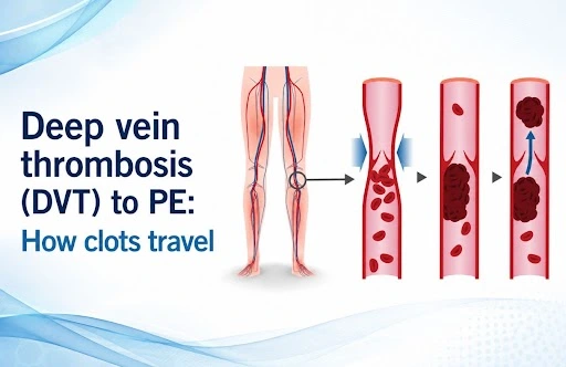
A Deep vein  thrombosis becomes a pulmonary embolism when a piece of the clot breaks free and travels through the bloodstream.

The general route looks like this:

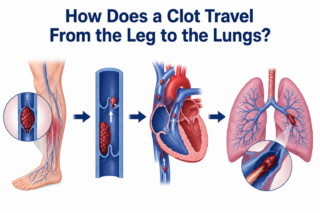
Deep leg veins → inferior vena cava → right atrium → right ventricle → pulmonary arteries → lungs

Once a fragment of the clot detaches, it’s called an embolus. It moves through the venous system toward the right side of the heart and from there into the pulmonary circulation. If it becomes lodged in a pulmonary artery, that’s a pulmonary embolism.

How serious the resulting PE is depends largely on the embolus’s size, where it lodges, and how much cardiopulmonary reserve the patient has. A large clot can meaningfully disrupt blood flow through the lungs and put real strain on the right side of the heart.

Also read: Deep vein thrombosis treatment | Cardiologist in Hyderabad 

Why Early DVT Treatment Matters

Once DVT is diagnosed or strongly suspected, the treatment goal is to stop the clot from growing and to lower the risk that a piece of it breaks away.

Anticoagulant medication is the backbone of DVT treatment. These drugs don’t dissolve the existing clot directly, but by limiting further clot formation, they give the body time to break down the thrombus on its own.

The specific anticoagulation plan, which drug and for how long,, is tailored to the individual, taking into account:

  • Where the clot is and how extensive it is
  • The patient’s bleeding risk
  • Any history of prior DVT or PE
  • Whether cancer or another underlying condition is driving clot formation
  • Recent surgery, hospital stays, or long periods of immobility
  • Other relevant health conditions

In other words, treatment isn’t one-size-fits-all just because a clot is present; it has to match the patient’s broader clinical picture.

Recognizing When Deep vein thrombosis Has Become PE

Pulmonary embolism should be on the radar any time a patient with known or suspected DVT suddenly develops heart or lung symptoms.

Typical warning signs of PE include:

  • Sudden or unexplained shortness of breath
  • Chest pain that worsens with deep breaths
  • A racing or irregular heartbeat
  • Persistent cough
  • Coughing up blood
  • Dizziness or fainting

None of these symptoms is unique to PE on its own, which is exactly why proper clinical evaluation and testing are essential rather than guesswork.

A typical scenario: someone recovering from a long period of immobility first notices swelling in one calf. If they then develop sudden breathlessness and sharp chest pain that worsens with breathing, that combination should prompt urgent evaluation for DVT-related PE.

How Pulmonary Embolism Is Treated

The right approach to pulmonary embolism treatment depends on how sick the patient is, how much of the pulmonary circulation is blocked, and the risk that their condition could worsen.

Anticoagulation remains the mainstay for preventing further clotting. For patients who are hemodynamically unstable or have a more severe PE, more aggressive interventions may be warranted depending on the clinical picture and the resources available.

Diagnostic workup commonly includes:

  • D-dimer testing in the right clinical context
  • Compression ultrasound to check for DVT
  • CT pulmonary angiography to confirm PE
  • ECG and cardiac biomarkers to assess risk
  • Echocardiography in select cases, to check how the right side of the heart is coping

The goal isn’t just to find the clot  it’s to understand how dangerous it is and how urgently the patient needs treatment.

When to Involve a Pulmonary Embolism Specialist

A pulmonary embolism specialist becomes especially relevant in complex cases: an extensive PE, significant strain on the heart and lungs, or a situation where advanced procedures like catheter-based clot removal are being considered.

For patients or referring doctors searching for a top pulmonary embolism doctor, a pulmonary embolism specialist in India, or a pulmonary embolism doctor nearby, it’s worth prioritizing a clinician experienced specifically in venous thromboembolic disease, including catheter-based and multidisciplinary treatment options where they apply.

For clinicians, the key takeaway is that DVT and PE aren’t two unrelated diagnoses; they’re two stages of the same underlying disease process, venous thromboembolism, and should be assessed together rather than in isolation.

The DVT-to-PE Pathway, Step by Step

  1. A clot forms in a deep vein.
  2. Part of the clot breaks loose.
  3. The embolus travels through the venous system.
  4. It reaches the right side of the heart.
  5. It passes into the pulmonary arteries.
  6. It may block blood flow through the lungs.
  7. The result is a pulmonary embolism.

This chain of events is exactly why catching DVT early and treating it properly matters so much in preventing a potentially life-threatening complication.

Also read : Pulmonary Embolism in India: Emergency & Finding Specialist 

When to Seek Specialist Care Urgently

Sudden breathlessness, unexplained chest pain, coughing up blood, fainting, or a noticeably fast heart rate in someone with known or suspected DVT calls for immediate medical attention. PE can be life-threatening, and it’s not something to wait out or try to manage without professional evaluation.

For patients dealing with complex DVT or pulmonary embolism, Dr. C. Raghu offers specialized cardiovascular care, including catheter-based treatment for venous thrombosis and pulmonary vascular disease.

If you or someone you know has suspected or confirmed DVT or pulmonary embolism, consult Dr. C. Raghu for a specialist evaluation and a treatment plan suited to your specific condition.


Dr raghu logo


Dr raghu logo


Call us now if you are in a medical emergency need, we will reply swiftly and provide you with a medical aid.


For Appointment with Dr Raghu: +91 80085 36699
Dr Raghu’s Patient Coordinator: +91 72860 10203
Dr Raghu’s Office Experience: +91 90001 65962
Dr Raghu’s Social Media Communication Channel: +91 97046 51708

Dr. Raghu | Heart Specialist in Hyderabad
Yashoda Hospitals, Raj Bhavan Road, Somajiguda, 1st floor, Room No. 115, Hyderabad -500082, Telangana

Best Cardiologists in Hyderabad


© 2024, Dr. Raghu. All rights reserved. Design & Developed by AMSDigital.in

[gtranslate]
+91-9542475650