
Best Heart Doctor in Hyderabad Why Dr. C Raghu
Best heart doctor Comprehensive Cardiac Care — Why It Matters for the Patient Cardiac conditions do not present in isolation.
Best heart doctor Comprehensive Cardiac Care — Why It Matters for the Patient Cardiac conditions do not present in isolation.
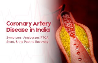
Coronary artery disease (CAD) kills more Indians than any other single condition. It strikes earlier in the Indian population than

India’s Coronary Disease Crisis — and Why the Quality of the Doctor Matters Angioplasty expert in India Coronary artery disease
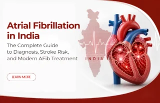
The Most Common Heart Rhythm Disorder and Why Knowing About It Protects Your Brain Atrial fibrillation treatment (AFib or AF)
The Heart Valve Condition That Affects Millions — and the Procedure Changing Its Outcome Severe aortic stenosis is one of

When a patient or family searches for a top cardiologist in Hyderabad, they often arrive at aggregator lists—rankings assembled from

Heart Attack Crisis — and Why Every Family Needs to Understand It Heart Attack Causes is the leading cause of

cardiologist in Hyderabad In today’s digital world, long hours of sitting have become a normal part of life—whether it’s working

Many people worry about sudden chest pain or breathlessness. They want clear steps to cut their heart risk. Recent research

Handle Cardiac Emergencies Many people face sudden chest pain or breathlessness at home. Families often do not know what to

CHIP Angioplasty Heart disease doesn’t always come with a warning. Many people discover they have coronary artery disease only after symptoms

When the Heart Speaks, Are You Listening? Heart valve You are the kind of person who reads nutrition labels. You
© 2024, Dr. Raghu. All rights reserved. Design & Developed by AMSDigital.in
+91 95424 75650
For Appointment with Dr Raghu:
+91 80085 36699
Dr Raghu’s Patient Coordinator:
+91 72860 10203
Dr Raghu’s Office Experience:
+91 90001 65962
Dr Raghu’s Social Media Communication Channel:
+91 97046 51708
