Mitral Valve Stenosis : Symptoms, Diagnosis, Treatment
What is Mitral Valve Stenosis?
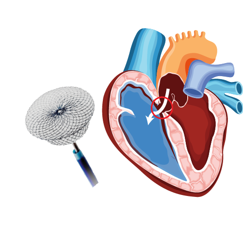
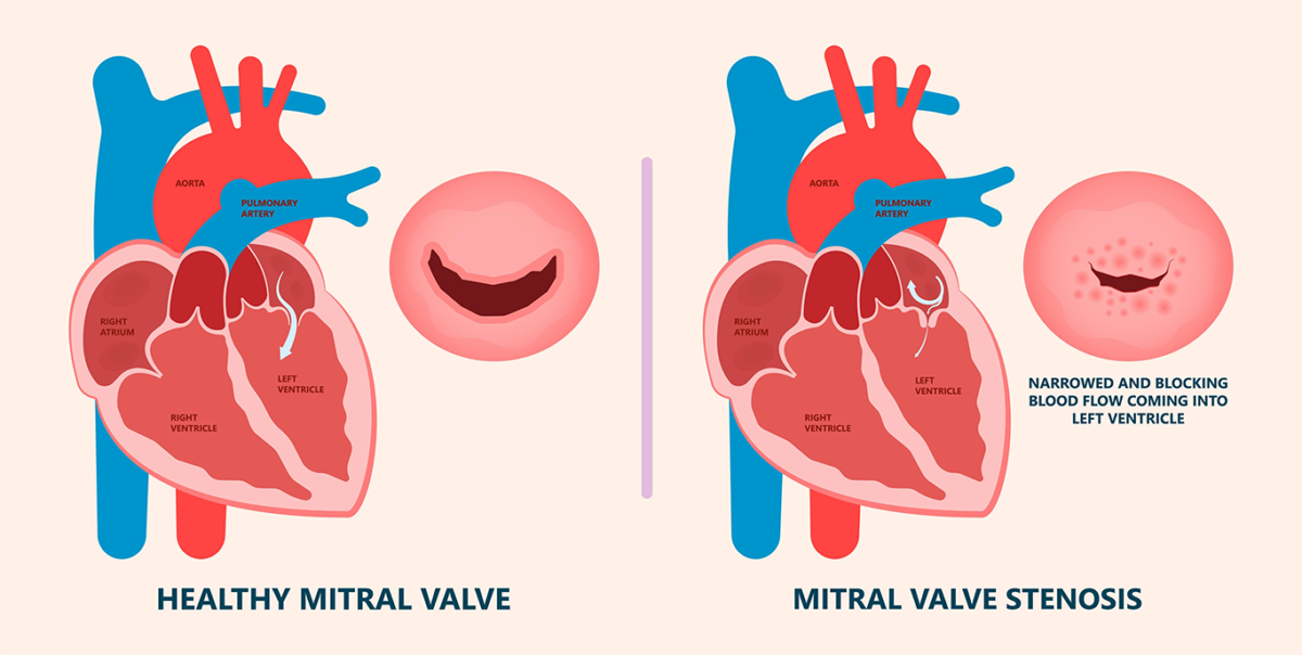
- The narrowing of the mitral valve, which lies between the upper left atrium and lower left ventricle, is called mitral valve stenosis.
- This condition limits the blood flow from the left atrium to the left ventricle and causes backflow of blood in the left atrium, blood vessels of lungs and right side of the heart.
- Symptoms do not develop until 10 to 20 years after stenosis progression. But in India and other developing countries, progression is very rapid and develops within 6 to 8 years only.
- The severity of this condition can be graded from mild to severe.
What are the causes of mitral valve stenosis?
- Rheumatic fever, a complication of a Streptococcus infection
- Calcium deposits due to an older age
- Congenital defect of mitral valve
- Autoimmune disease such as lupus
- Radiation therapy to the chest
What are the symptoms of mitral valve stenosis?
- Shortness of breath
- Cough
- Wheezing
- Abnormal heart rhythms (Arrhythmias)
- Rapid heartbeats (Palpitations)
- Chest pain or discomfort
- Heart murmurs
- Fatigue
- Swelling in legs, feet, or ankle
What are the complications of mitral valve stenosis?
- Heart enlargement: Enlargement of the upper left chamber (left atrium) and right side of the heart due to pressure build in mitral valve stenosis.
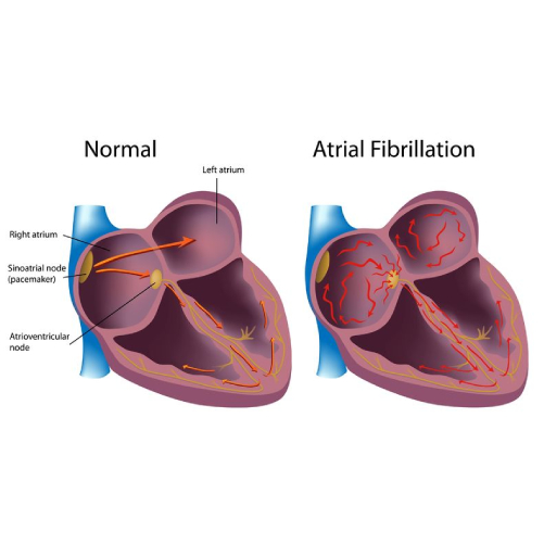
- Atrial fibrillation: Stretching and enlargement of upper chambers (atrium) can result in abnormal heart rhythm.
- Blood clots: If the abnormal or rapid heartbeat is left untreated, blood clots form in the left atrium. These clots can break and travel to other parts of the body resulting in serious complications such as paralytic stroke.
- Heart failure: Pressubuildsild up in the upper left chamber and lungs results in fluid accumulation resulting in heart failure.
- Pulmonary arterial hypertension: Increased blood pressure in the arteries that carry blood from the heart to lungs (pulmonary arteries).
- Stroke
- Endocarditis (infection of the heart)
How is mitral valve stenosis diagnosed?
Mitral valve stenosis is undiagnosed until a few years after stenosis progression. If the doctor thinks he or she might have the disease, the followings tests are performed.
- Physical examination: To check the presence of heart murmurs.
- Electrocardiogram: To check the electrical activity of the heart.
- Echocardiogram: Utilizes ultrasound images to examine the heart’s chambers, valves and blood vessels.
- Trans-oesophagal echocardiogram (TEE): This test helps to determine the cause and severity of the stenosis.
- Cardiac catheterization: To perform coronary angiography before mitral valve replacement surgery in patients for more than 40 years.
- Chest X-ray
How is mitral valve stenosis treated?
Treatment of mitral valve stenosis depends upon the symptoms and severity.
- If the symptoms are mild, one should probably able to do daily activities and mild exercise and if the symptoms are moderate to severe, should avoid intense exercise
- Limit salt in the diet
- Anti-coagulation therapy to prevent blood clot formation
- Medications that keep control over heartbeat and rhythm
- Open heart surgery for mitral valve replacement
- Percutaneous balloon valvotomy: Catheter is passed through blood vessels of heart, and the balloon is inflated to widen or open the mitral valve to correct the blood flow.
What is the uniqueness of treatment of mitral valve stenosis by Dr. C Raghu?
- Decision making in mitral stenosis is crucial to determine whether surgical valve replacement is needed or a non-surgical balloon mitral valvotomy be performed. This is eminently done by Dr. Raghu with vast clinical experience.
- Focussed echocardiography and Transesophageal echocardiography need to be performed for appropriate decision making. Once again Dr. Raghu is personally involved in this decision making a journey.
Being an expert in Interventional Cardiology has performed to date more than 400 balloon mitral valvotomy procedures.
Our Specialities
- Conditions
Conditions
- Acute limb ischemia
- Chronic limb ischemia
- Aortic stenosis
- Mitral valve stenosis
- Mitral valve regurgitation
- Atrial fibrillation
- Tachycardia
- Bradycardia
- Palpitations
- High blood pressure
- Atrial septal defect
- Ventricular septal defect
- Patent ductus arteriosus
- Cardiac amyloidosis
- Hypertrophic cardiomyopathy
- Varicose veins
- Deep vein thrombosis (DVT)
- Myocarditis
- Endocarditis
- Pericarditis
- Peripheral arterial disease
- Pulmonary artery hypertension
- Pulmonary embolism
- Cath lab procedures:
Cath lab procedures:
- Coronary Angiogram
- Primary Angioplasty
- Coronary Angioplasty
- CHIP Angioplasty
- Aortic valve replacement surgery
- Mitral valve replacement surgery
- Device closure for Atrial septal defect
- Device closure for Ventricular septal defect
- Device closure for Patent Ductus Arteriosus
- Transcatheter aortic valve replacement (TAVR)
- Inferior vena cava (IVC) filter
- LA appendage closure
- Fistuloplasty
- Balloon mitral valvotomy
- 24 hours emergency services
24 hours emergency services
- Clinics- weekly basis/monthly basis/ Yearly basis
Clinics- weekly basis/monthly basis/ Yearly basis
- Prevention of cardiovascular diseases
Prevention of cardiovascular diseases
- Diagnosis
Diagnosis
BOOK AN APPOINTMENT

Dr. RAGHU | Mitral Valve Stenosis Treatment
MD, DM, FESC, FACC, FSCAI
Cardiology Coronary, Vascular and
Structural Interventions
Cardiology Coronary, Vascular and
Structural Interventions
Conditions & Diseases
Angioplasty
Aortic Stenosis
Atrial Fibrillation
