Ventricular Septal defect – Symptoms, Causes & Closure in Hyderabad
What is Ventricular septal defect?
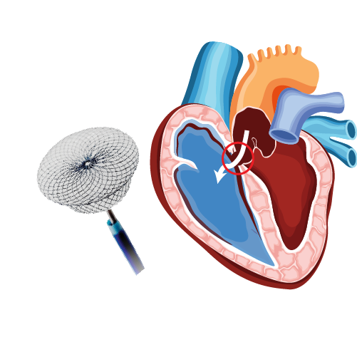
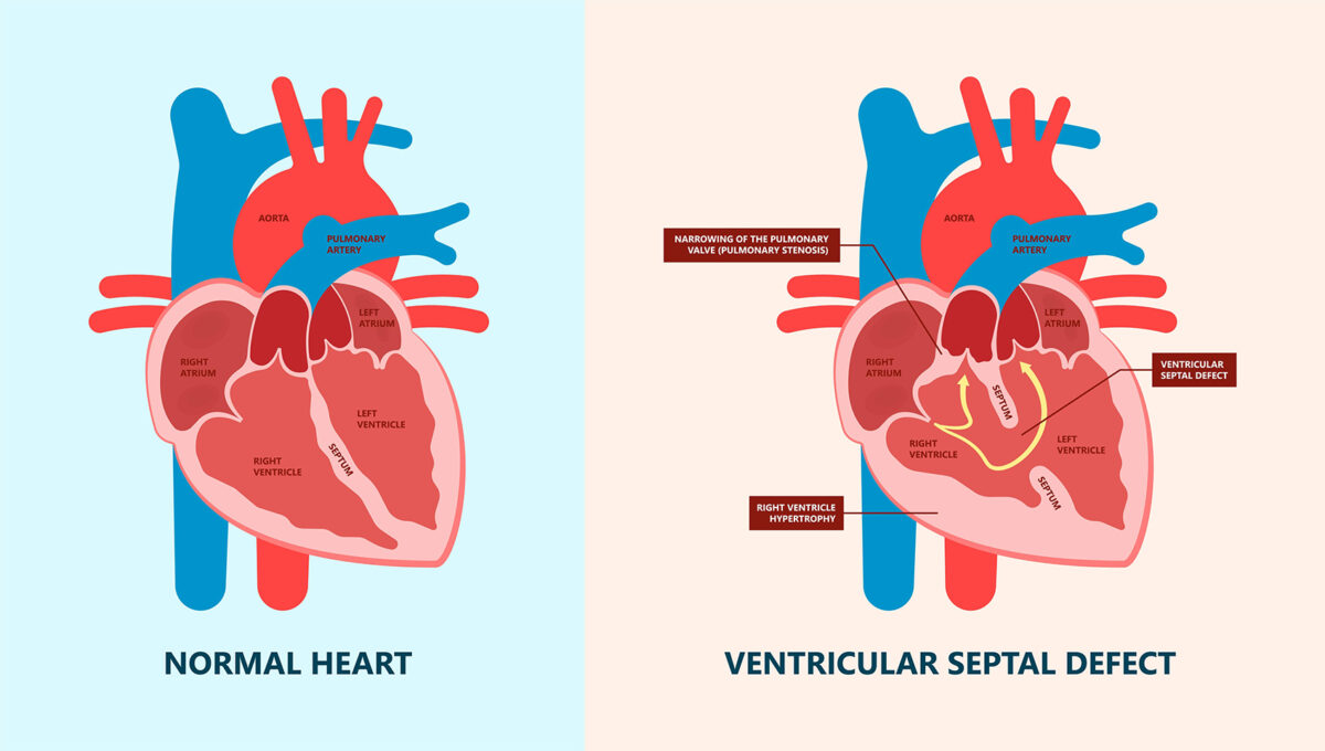
- Ventricular septal defects (VSD) are congenital abnormalities characterized by a structural deficiency of the ventricular septum that separates lower chambers of the heart.
- Due to the presence of a hole in the ventricular septum, blood from the left ventricle mixes with that in the right ventricle which increases the pressure and eventually weakens the pumping ability of the heart.
- Types of VSD – Atrioventricular canal type, Muscular, Membranous, Conoventricular, Canal septal or right ventricular outlet.
- 80 % of VSD type is membranous type.
- Small ventricular defects have no major problems and close by itself. Whereas, medium or large defects require surgery to prevent complications.
What are the symptoms of ventricular septal defect?
- If the ventricular defect is small, symptoms may not appear until later stages of childhood. Whereas, for the greater defects following symptoms will be presented.
- Rapid breathing or breathless
- Decreased appetite – failure to thrive
- Frequent tiredness
- Murmurs
- Rapid or irregular heartbeat
What are the causes of ventricular septal defect?
- Congenital heart diseases are due to problem in heart development
- Genetics
- Environmental factors
What are the risk factors of ventricular septal defect?
- A genetic problem such as Down’s syndrome
What are the complications of ventricular septal defect?
- Heart failure: This condition will develop in case of medium to large VSD because the heart needs to work harder to pump enough blood to the body.
- Pulmonary hypertension: increased blood flow to the lungs due to VSD.
- Endocarditis
- Other heart problems such as rhythm abnormalities, and valve problems
How are ventricular septal defects diagnosed?
- Chest X-ray
- Electrocardiogram
- Echocardiogram – identifies anatomy of the defect and shut size.
- Cardiac catheterization: Examines the heart structures, blood pressure and blood oxygen levels in its chambers.
Why should we close VSD?
- Most VSD may be closed in childhood
- Large non-restrictive VSD present in infancy– need early surgery
- Smaller restrictive defects can remain relatively asymptomatic but can result in long-term sequelae such as pulmonary hypertension, left heart dilation, arrhythmia, aortic regurgitation, double chamber right ventricle, or endocarditis
- VSD persist beyond age 8 years – does not close
- Hemodynamically insignificant VSDs can lead to the risk of developing endocarditis.
Which patients need closure?
- Cardiomegaly or left heart dilation by echocardiogram
- Qp: Qs greater than 1.5
- Failure to thrive
- Worsening New York Heart Association class symptoms
- Recurrent respiratory infections
- History of infective endocarditis
What are the contraindications of device closure?
- Irreversible pulmonary vascular disease
- Patients with antiplatelet therapy
- Active infection or sepsis
- Anatomical concerns:
- Inadequate rim (<2mm) below the aortic valve in a membranous VSD
- Supracristal defects
- Aortic valve cusp prolapse
- Misalignment VSDs
Why device closure and not surgery?
- Avoids open heart, on bypass pump surgery
- Short hospital stay (< 3 days)
- Pain and delayed recovery with surgery
- Cosmetic and stigma consequences of surgery
How is VSD treated?
- Initially, the child is given anaesthesia for sedation.
- Small incision or cut is made in the groin area and catheter will be inserted and directed towards the ventricular septum between the lower chambers of the heart.
- Then, a device to occlude the defect is guided with catheter towards defect.
- Amplatzer ventricular septal occluder is used in ventricular septal defect closure :
- These are self-expandable, double-disc device
- This is made from nitinol wire mesh
- This can be ruptured and redeveloped for precision in placement
- This device provides complete closure of the defect
- Once the device is in the right position, it is deployed between the two discs to occlude the defect and catheter will be removed.
- Vitals such as Blood pressure, heart rate, temperature, respiration rate, blood oxygen level will be monitored.
- Blood tests, Chest X-ray and Electrocardiogram(ECG), Echocardiogram will be done
- Infections and bleeding manifestations are checked at the insertion site
How to prevent VSD?
In most cases, you cannot prevent congenital abnormalities. However, the following precautions are taken.
- Early prenatal checkup
- Balanced diet
- Regular exercise
- Avoid alcohol, tobacco, and illegal drugs
- Control sugar levels
What is the uniqueness of VSD closure by Dr. Raghu?
Dr. Raghu team has performed date 100 VSD device closure procedure. One of the early adaptors to perform VSD device closure in India, Dr. Raghu, has organized the first-ever workshop on VSD device closure in 2011.
Our Specialities
- Conditions
Conditions
- Acute limb ischemia
- Chronic limb ischemia
- Aortic stenosis
- Mitral valve stenosis
- Mitral valve regurgitation
- Atrial fibrillation
- Tachycardia
- Bradycardia
- Palpitations
- High blood pressure
- Atrial septal defect
- Ventricular septal defect
- Patent ductus arteriosus
- Cardiac amyloidosis
- Hypertrophic cardiomyopathy
- Varicose veins
- Deep vein thrombosis (DVT)
- Myocarditis
- Endocarditis
- Pericarditis
- Peripheral arterial disease
- Pulmonary artery hypertension
- Pulmonary embolism
- Cath lab procedures:
Cath lab procedures:
- Coronary Angiogram
- Primary Angioplasty
- Coronary Angioplasty
- CHIP Angioplasty
- Aortic valve replacement surgery
- Mitral valve replacement surgery
- Device closure for Atrial septal defect
- Device closure for Ventricular septal defect
- Device closure for Patent Ductus Arteriosus
- Transcatheter aortic valve replacement (TAVR)
- Inferior vena cava (IVC) filter
- LA appendage closure
- Fistuloplasty
- Balloon mitral valvotomy
- 24 hours emergency services
24 hours emergency services
- Clinics- weekly basis/monthly basis/ Yearly basis
Clinics- weekly basis/monthly basis/ Yearly basis
- Prevention of cardiovascular diseases
Prevention of cardiovascular diseases
- Diagnosis
Diagnosis
BOOK AN APPOINTMENT

Dr. RAGHU
MD, DM, FESC, FACC, FSCAI
Cardiology Coronary, Vascular and
Structural Interventions
Cardiology Coronary, Vascular and
Structural Interventions
Conditions & Diseases
Angioplasty
Aortic Stenosis
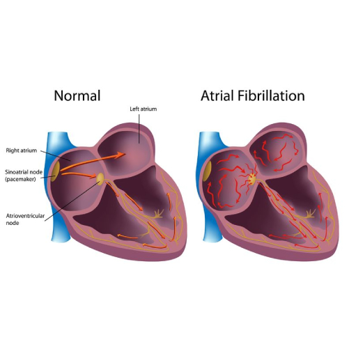
Atrial Fibrillation
