Coronary Angiogram
CORONARY ANGIOGRAM
What is Coronary Angiogram?
Coronary Angiogram is a diagnostic procedure that uses X-ray imaging to identify narrowing of the blood vessels supplying the heart.
- Coronary arteries are blood vessels supplying blood to the heart
- A contrast agent (iodine dye) is injected through a long thin plastic tube (catheter),which will demarcate the heart vessels and narrowing.
- A coronary angiogram is also called coronary angiography (CAG), coronary arteriography (CART).
- In certain countries, these are part of the general group of procedures known as cardiac catheterization.
- It is a gold standard procedure for diagnosing blockages in the arterial system.
How does one reach the heart during a coronary angiogram?
To identify any heart vessel blocks the catheter is inserted majorly from two different sites.Groin (trans-femoral angiogram)
- Wrist (trans-radial angiogram)
- In extremely rare circumstances based upon the clinical scenario alternate access sites can also be used.
When is coronary angiogramadvised?
History of new-onsetunusual chest pain
- Abnormal noninvasive cardiac stress test results
- Abnormal CT angiogram results
- In the setting of heart attack
- People with poor left ventricular function
- Preoperative evaluation before major heart surgery like valve replacement, closure of heart defects, tumour removal etc.
- To evaluate the functioning of stent or bypass graft after surgery
- As a part of the preoperative workup for certain high-risk surgeries such as vascular surgery, aortic surgery etc.
What are the risks of a coronary angiogram?
Generally, this is procedure is safe and have no major complications. However, some potential risks and complications include:
- Allergic reaction to the contrast dye
- Kidney damage consequent to contrast dye utilization
- Infection and bleeding or burning at the site of the procedure
- Blood clots formation potentially leading to a heart attack or stroke
- Irregular heart rhythms (arrhythmias)
- Leakage from the artery leading to a pseudoaneurysm
Understanding the procedure
Before the procedure
Dietary instructions:
- Stop heavy meals or processed food before 8 hours of the procedure
- Stoplight meals, milk, carbonated drinks before 6 hours
- Stop drinking water or clear fruit juice before 2 hours
- Adequate hydration is important to reduce the risk of kidney damage
General instructions:
List all the current medications and supplements used
- Any allergies towards the medications should be reported
- Vitals are checked
- Baseline ECG, echocardiography, blood investigations to estimate kidney function and haemoglobin are essential before an angiogram
- Reports of previous blood investigation reports and medical records should be available
- It is important to have some beside you to take care of you and to drive back home after the procedure
- Plan to have someone beside you to take care of you and back home after the procedure
During the procedure:
To reduce the risk of infection the following precautions are taken
- Hand wash and sanitization of the team of doctors
- Your skin will be washed with disinfectant
- Hair is clipped at the site of catheter insertion
- You will be made to lie on the X-ray table and strapped
- An intravenous cannula will be inserted
- Electrodes will be placed over your chest to monitor for vitals during the procedure
- A mild sedative will be administered to make you drowsy but still alert.
- Local anaesthesia at the catheter insertion site to make the area numb.
- A catheter is inserted with guidewire support into the radial/femoral artery and advanced under X-ray guidance to reach the heart.
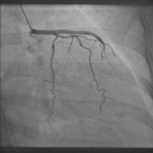
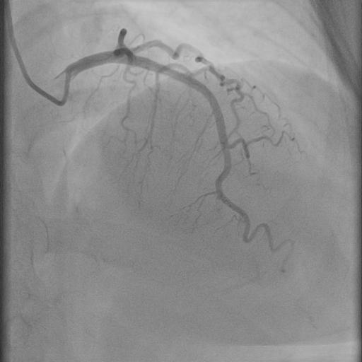
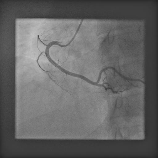
- Medicine to opacify the heart vessels (contrast dye) will be injected and X-ray is taken.
- Opacification of the heart vessels identifies the blocks
- The heart has three blood vessels, on the left the main vessel ( left main coronary artery -LMCA) that divides into a left anterior descending coronary artery (LAD) and leftcircumflex coronary artery (LCX); Right coronary artery (RCA) on the right.
- The number of blood vessels affected, lesion- number location, and characteristics are assessed
- Once the procedure is completed, the catheter is removed and a bandage is placed over the procedure site.
After the procedure
Vitals such as Blood pressure, heart rate, temperature, respiration rate, blood oxygen levels will be monitored.
- One needs to rest for 6 to 8 hours.
- If the catheter insertion is at the groin, you may be asked not to bend or cross your legs for 12 hours.
- Infection and bleeding are checked for at the procedure site
- It is very important to take plenty of liquids that keep you hydrated as well as wash off contrast from the body. Accumulation of contrast leads to kidney damage.
- Blood investigations and ECG need to be repeated before the follow-up visit

How does catheter coronary angiogram compare with CT coronary angiogram?
| Catheter coronary angiogram | CT coronary angiogram |
| A catheter is inserted through an artery to reach the heart and directly inject contrast to opacify the heart vessels | An intravenous cannula is inserted and contrast material injected which passes through the veins to reach the heart vessels |
| Advantages: | Advantages: |
| The gold standard for delineating blocks in the heart vessels | Ideal procedure to rule out blocks in the heart vessels |
| Allows performing treatment, angioplasty-stent, if required procedure due can be performed with a limited quantity of contrast | Ideal procedure to rule out blocks in the heart vessels |
| Essential for planning complex angioplasty | Very useful for people witha low probability of heart vessel block |
| Mandatory before bypass surgery | Identify small-sized arteries that may not be visualized in a scenario of totally occluded heart vessel |
| Enables blood pressure measurement in various chambers of the heart; allows one to understand heart functioning ability | Useful for delineating bypass graft vessels |
| Does not require a hospital stay | |
| Limitations: | Limitations: |
| Invasive procedure | Limited information about heart vessel narrowed lesions |
| Remote risk of brain stroke and heart attack | Cannot plan or perform angioplasty-stent or bypass surgery based on CT angiogram results |
| Complications at the site of catheter insertion | Requires a relatively larger volume of contrast that can potentially worsen kidney function in vulnerable patients |
| Allergic reaction to contrast | Allergic reaction to contrast material |
What is the uniqueness of the angiograph procedure by Dr C Raghu?
Our team is a pioneer in performing angiography, angioplasty procedure through the wrist. Procedures performed through the wrist have a zero per cent of bleeding complications from the catheter insertion site. Over the last 20 years, Dr C Raghu’s team has performed 40 thousand procedures through the wrist with high success and low complication rates.
- Conditions
- Acute limb ischemia
- Chronic limb ischemia
- Aortic stenosis
- Mitral valve stenosis
- Mitral valve regurgitation
- Atrial fibrillation
- Tachycardia
- Bradycardia
- Palpitations
- High blood pressure
- Atrial septal defect
- Ventricular septal defect
- Patent ductus arteriosus
- Cardiac amyloidosis
- Hypertrophic cardiomyopathy
- Varicose veins
- Deep vein thrombosis (DVT)
- Myocarditis
- Endocarditis
- Pericarditis
- Peripheral arterial disease
- Pulmonary artery hypertension
- Pulmonary embolism
- Cath lab procedures:
- Coronary Angiogram
- Primary Angioplasty
- Coronary Angioplasty
- CHIP Angioplasty
- Aortic valve replacement surgery
- Mitral valve replacement surgery
- Device closure for Atrial septal defect
- Device closure for Ventricular septal defect
- Device closure for Patent Ductus Arteriosus
- Transcatheter aortic valve replacement (TAVR)
- Inferior vena cava (IVC) filter
- LA appendage closure
- Fistuloplasty
- Balloon mitral valvotomy
- 24 hours emergency services
- Clinics- weekly basis/monthly basis/ Yearly basis
- Prevention of cardiovascular diseases
- Diagnosis
BOOK AN APPOINTMENT
Coronary Angiogram PART-1
Coronary Angiogram PART-2
+91 95424 75650

© 2024, Dr. Raghu. All rights reserved. Design & Developed by AMSDigital.in