
mitral-balloon.jpg

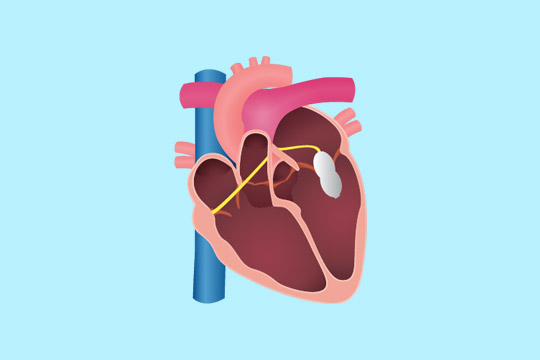
Mitral stenosis is the narrowing of the valve present between the left chambers of the heart (mitral valves), thereby blocking the blood flow. This condition usually develops several years after a person had a rheumatic fever. Mitral stenosis is treated with balloon valvotomy, when the medications do not reduce the symptoms. If left untreated, mitral stenosis can result in various heart complications.

Mitral valvotomy (or valvuloplasty), also known as percutaneous balloon dilation, is a minimally invasive procedure that involves widening a mitral valve using a balloon catheter, a thin, flexible tube with a balloon at the tip. This procedure improves the overall function of the heart.

What are the risks of mitral valvuloplasty?

The risks associated with percutaneous balloon dilation include:

  • Blood clot formation or tears in the heart
  • Backward flow of the blood (mitral valve regurgitation) due to damaged valve
  • Restenosis of the mitral valve

What happens before the procedure?

Your doctor will explain the procedure in detail and provide you the opportunity to ask any questions; do not hesitate to ask any questions related to the procedure.

Your healthcare team will give you certain instructions to prepare for the procedure:

  • You will be asked not to eat or drink anything after midnight, on the previous night of the procedure; you can drink water up to 4 hours of the procedure.
  • A blood test may be done to evaluate the time required for the blood to clot; other blood tests may also be performed.
  • You may be asked to stop taking certain medicines that may involve with the blood clotting process.

Notify your doctor if you:

  • Had an allergic reaction to any contrast dye, iodine or seafood.
  • Are sensitive to any medications, tape, latex, or anaesthetic agents.
  • Have a pacemaker.
  • Have any body piercings on the abdomen or chest.
  • All the medications you are taking, including over-the-counter drugs, herbal supplements, blood thinners, etc.
  • Have heart valve disease, as antibiotic drugs may be given before this procedure.
  • Have a history of bleeding disorders
  • Are or may be pregnant

Before the procedure, the area near the catheter insertion site (the groin area) may be shaved. Your physician may order other preparations for the procedure based on your medical condition.

How is mitral valvuloplasty performed?

On the day of the procedure, you should remove your jewellery and other objects that may interfere with the procedure. You will be asked to change into a hospital gown and empty your bladder before the procedure.

An intravenous (IV) line will be attached to your arm or hand to inject medications and administer IV fluids, if needed. You may receive a sedative to help you relax.

The following are the steps generally involved in a balloon valvuloplasty:

  • A local anaesthetic is injected at the insertion site.
  • Once the anaesthesia sets in, a sheath or an introducer (a plastic tube) will be inserted into the blood vessel.
  • A valvuloplasty catheter is inserted through the sheath into the blood vessel. The catheter is advanced through the aorta into the heart valve. The catheter may be guided by a fluoroscopy (a special x-ray).
  • Once the catheter reaches the precise location, a contrast dye is injected into the valve to get a clear image of the area.
  • The balloon is then inflated and deflated several times.
  • The catheter is then removed.
  • The catheter insertion site is closed using a sterile bandage.

What happens after the procedure?

After the procedure, you will be moved to the recovery room. You should remain flat on bed for several hours after the procedure. Your vital signs, and circulation and sensation in the affected arm or leg, the insertion site will be monitored regularly.

Medicines may be given for pain or discomfort near the insertion site. You will be asked to drink water and other fluids to eliminate the contrast dye from your body. You can return to your regular diet after the procedure.

Mostly, you may have to spend the might in the hospital, based on your medical condition and recovery.

Your healthcare team will give you instructions to be followed after leaving the hospital:

  • Keep a check on the insertion site for unusual pain, bleeding, swelling, or discoloration.
  • Keep the insertion site clean and dry.
  • Do not participate in any strenuous activities. Your doctor will inform you when you can resume normal activities and return to work.

Call your doctor immediately if you have any of the following:

  • Fever or chills
  • Severe pain, swelling, redness, bleeding or other leakage from the insertion site
  • Numbness, coolness or tingling sensations in the affected extremity
  • Pain or pressure in the chest, nausea or vomiting, sweating, or dizziness
  • Reduced urination
  • Swelling of the abdomen or extremities
  • Over 3 pounds weight gained in a day


cci-dialysis_fistuloplasty.jpg

January 16, 2021 Dr. RaghuHearty Life

Hemodialysis is a preferred treatment option for patients with chronic kidney disease. In this t procedure, the blood is filtered outside the body by a dialyzer or “artificial kidney”. For hemodialysis, a vascular access is created to insert the needles that connect the dialyzer, thereby allowing the blood to move out and return to the body at a high rate. An arteriovenous fistula is the preferred vascular access for hemodialysis.

An arteriovenous (AV) fistula is created by connecting an artery to a vein, usually in the wrist or upper arm. But sometimes, the fistula can become infected, blocked or narrowed. The blocked fistula can be treated by a balloon fistuloplasty.

arteriovenous (AV) fistula

What is balloon fistuloplasty?

Balloon fistuloplasty is a procedure in which any blockage or narrowing in the fistula is located by using a dye, and the blockage is relieved by stretching the blood vessels with a special balloon.

In this procedure, a small balloon is inflated for several times at the narrowed regions of a fistula; if required a stent may also be placed. This technique widens the lumen and facilitates the process of dialysis.

Why perform fistuloplasty?

A fistula can age and cause problems like clotting and scarring, thereby decreasing its function and effectiveness of dialysis. Clots can decrease the rate of blood flow or block the fistula completely. If these problems are left untreated, it may lead to the failure of fistula.

Thus, it is important to treat the narrowing or blockage at an early stage, so that the fistula works well, and dialysis occurs without any complications.

What are the risks of dialysis fistuloplasty?

Fistuloplasty is usually a safe procedure, but some complications may occur rarely. The common risks and complications associated with fistuloplasty include:

  • Bruising around the site of insertion of the needle, which may become large and uncomfortable (rarely).
  • Infection of large bruises, which may need antibiotic treatment or surgical intervention.
  • Allergic reaction to the dye, which may present as a skin rash
  • Circulatory problems due to the damage to the artery or fistula caused by catheter or balloon
  • Treatment failure, which will require surgical intervention

How to prepare for the procedure?

Your healthcare provider will give you instructions to prepare for the procedure, which may include:

  • You may be asked not to eat or drink anything for six hours before the procedure; you can drink water up to two hours before.
  • Some blood tests may be performed before the procedure to evaluate the risk of bleeding.
  • If you are diabetic, ask your doctor to alter the treatment regimen.
  • Ask your doctor which medicines you can continue to take, and which ones you should stop.
  • If you are taking any antiplatelets or anticoagulants, you may have to stop taking these medicines a few days before.
  • Ensure that you have an adult to drive you home and accompany you overnight.

Inform your healthcare provider if you:

  • Are allergic to iodine, or have any other allergies
  • Have a history of reaction to the dye used for CT scan or X-rays
  • May be or are pregnant
  • Are a diabetic

What happens during the procedure?

The following are the steps usually performed in a fistuloplasty:

  • You will be asked to lie on your back on an x-ray table. Some monitoring equipment will be attached to measure your heart rate and blood pressure.
  • The interventional radiologist will observe the fistula by using an ultrasound, which provides a clear picture of the fistula.
  • A small needle is inserted in the fistula, and a contrast dye is injected. This provides an image of the blood vessels, which helps to locate the narrowing or blockage.
  • A catheter (a small, flexible tube) with a balloon at the tip is then inserted in the blood vessel to reach the precise location.
  • Once the catheter reaches the site of the blockage or narrowing, the balloon is inflated and deflated several times from outside the body.
  • Sometimes, if the balloon does not improve the fistula, a permanent stent may be used to widen the narrowing.
  • Then the catheter is removed from the blood vessel, and the puncture site is stitched to prevent bleeding.

Usually, fistuloplasty takes about one hour, but the duration may vary in different patients.

What happens after the procedure?

You will have to stay in the hospital after the procedure for three to four hours for observation. Your pulse, blood pressure and oxygen levels in the body will be monitored regularly. You may return to your normal diet. The fistula is ready to use immediately after the procedure.

Your nurse will tell you when you can go home. You will need a friend or a family member to drive you home; using public transport is not recommended.

Your fistula should be ready for use immediately after the procedure.

What measures do I take after going home?

The following measures will help you recover better after a fistuloplasty:

  • Rest well on the day of the procedure and the next day. Then, you can return to your normal activities.
  • You can follow your normal diet.
  • Take the pain killers as prescribed r instructed.
  • Continue to take you regular medicines, as prescribed.
  • Do not take metformin until two days after the procedure.
  • You can have a bath or shower the next day.

Call your doctor immediately

if you have any of the following symptoms:

  • A lot of swelling and bruising
  • Severe pain at the puncture site that does not get better with painkillers
  • Bleeding at the puncture site
  • Change in the colour of your arm
  • Fever or chills
  • A lump, pus or discharge at the puncture site
  • Difficulty breathing or chest pain


diagnosis-procedure-cardiac_catheterization.jpg

Cardiac catheterization is a minimally invasive procedure used to detect and treat cardiovascular diseases. This procedure involves the insertion of a catheter (a thin hollow tube) into the large arteries or veins present in the neck, arm or groin, which is then guided to the heart using a special X-ray.


cci-cardiac_catheterization.jpg

January 16, 2021 Dr. RaghuHearty Life

Peripheral arteries are the blood vessels that deliver blood to the lower limbs. When the cholesterol accumulates in these blood vessels, the blood flow to the lower limbs gets blocked. Peripheral angioplasty is a minimally invasive procedure. It is done to restore the blood flow by opening the blocked peripheral arteries.

peripheral angioplasty

When is peripheral angioplasty performed?

  • Peripheral angioplasty is mostly indicated in the treatment of Peripheral Vascular Disease (the circulatory disorder caused by the blocked or narrowed blood vessels outside the heart).
  • Peripheral artery disease is the common condition, in which peripheral angioplasty is recommended.

Are there any risks with peripheral angioplasty?

Peripheral angioplasty is associated with the following risks:

  • Breathing problems
  • Bleeding
  • Blood clots
  • Infection
  • Kidney damage
  • Damage to the blood vessel

How to prepare for the procedure?

Before initiating the procedure, a physical examination and imaging tests are done to determine the overall health condition. Additionally, the following steps would help in a successful procedure and quicker recovery:

  • Inform the use of current medications, vitamin or mineral supplements.
  • Tell the doctor about any underlying disease condition.
  • Take the prescribed medicines.
  • Notify the doctor if you have any allergies.
  • Six to eight hours before the surgery do not eat or drink anything.

What happens during the procedure?

Local anaesthesia is administered in the upper thigh region. Once the anaesthesia sets, an incision is made on the upper thigh to insert the catheter. By using a high-resolution fluoroscopic, the catheter is guided to the blocked artery. When the catheter reaches the obstructed artery, the balloon is inflated to widen the blood vessel. Once the blood flow is restored, a stent is placed to prevent the risk of further blockage. Finally, the incision is closed and covered with a sterilized bandage.

What to expect after the procedure?

You will be placed in a recovery room and the vital parameters would be checked. For at least 3-6 hours, you need to remain still, to prevent bleeding from the incision site. Depending on the patient’s condition, the doctor will decide whether the person requires a hospital stay or not.
Before discharge, you will receive the following instructions:

  • Keep the wound clean and dry.
  • Do not lift heavyweights.
  • Avoid strenuous exercises for at least 24 hours after the procedure.
  • Drink plenty of fluids to help flush out the contrast dye from the body.

Care after peripheral angioplasty:

Although peripheral angioplasty clears the blockage, it does not treat the underlying cause of the blockage. So, to prevent the further risk of blockage, the following steps should be taken:

  • Maintain a healthy body weight.
  • Quit smoking.
  • Exercise regularly.
  • Take the prescribed medicines to prevent re-narrowing of the blood vessels.
  • Manage stress.
  • Avoid fatty-foods.
  • Have a low-salt and low-fat diet to prevent the risk of fluid retention.

Seek medical attention:

The following symptoms are the warning signs that require immediate medical attention:

  • Swelling in limbs
  • Chest pain
  • Shortness of breath
  • Fever associated with chills (over 101oF)
  • Weakness
  • Dizziness


disease-condtions-critical_limb_ischemia.png

January 16, 2021 Dr. RaghuHearty Life

Critical limb ischemia (CLI) refers to severe compromise of blood flow to alimb (hands or legs) which causes severe limb pain at rest or even loss of limb. It is the most advanced form of peripheral artery disease.

CLI numbers:

  • Prevalent in 2% patient over 70 years of age.
  • Within 1 year of diagnosis, 40-50% patientshave an amputation and 25% die.

How CLI leads to amputation?

Amputation occurs when there is marked ischemia of the limb owing to reduction of blood flow and increase demand in the limb.

Factors that reduce blood supply:

  • Diabetes mellitus
  • Severe Renal or Heart failure
  • Vasospastic diseases
  • Smoking

Factors that increase demand for blood flow:

  • Infection (cellulitis)
  • Skin breakdown
  • Trauma
  • Osteomyelitis

 Diagnosis of CLI:

Characteristic Duplex Ultrasound Digital – Subtraction Angiography Magnetic Resonance Angiography (MRA) Computed Tomographic Angiography (CTA)
Advantages
  • Noninvasive
  • Can Visualize & Quantitate severity
  • Gold Standard
  • High Resolution
  • Can guide intervention
  • Noninvasive
  • No radiation
  • No contrast
  • 3 D
  • Noninvasive
  • Higher Resolution than MRA
  • 3 D
Disadvantages
  • Operator dependent
  • Limited by
    dense calcification
  • Invasive
  • Radiation
  • Contrast
  • 2Dimensional
  • Lower Resolution than CTA
  • Claustrophobia
  • Contrast Image artifact if stent present
  • Radiation (25% of dose with DSA)
  • Contrast
  • Limited by calcification

Management plan for critical limb ischemia:

Critical Limb Ischemia Non-Healing Ulcer Rest Pain

MRA, CTA or invasive argiography shows lesion treatable by endovascular approach

  • Endovascular revascularization ->Wound care and atherosclerosis risk factor modification
  • Lesion treatable by open sugery at acceptable operative risk
    • Surgical revascularization ->Wound care and atherosclerosis risk factor modification
    • Consider primary amputation ->Wound care and atherosclerosis risk factor modification


Dr raghu logo


Dr raghu logo


Call us now if you are in a medical emergency need, we will reply swiftly and provide you with a medical aid.


For Appointment with Dr Raghu: +91 80085 36699
Dr Raghu’s Patient Coordinator: +91 72860 10203
Dr Raghu’s Office Experience: +91 90001 65962
Dr Raghu’s Social Media Communication Channel: +91 97046 51708

Dr. Raghu | Heart Specialist in Hyderabad
Yashoda Hospitals, Raj Bhavan Road, Somajiguda, 1st floor, Room No. 115, Hyderabad -500082, Telangana

Best Cardiologists in Hyderabad


© 2024, Dr. Raghu. All rights reserved. Design & Developed by AMSDigital.in

[gtranslate]
+91-9542475650