Aortic Disease | Dr Raghu

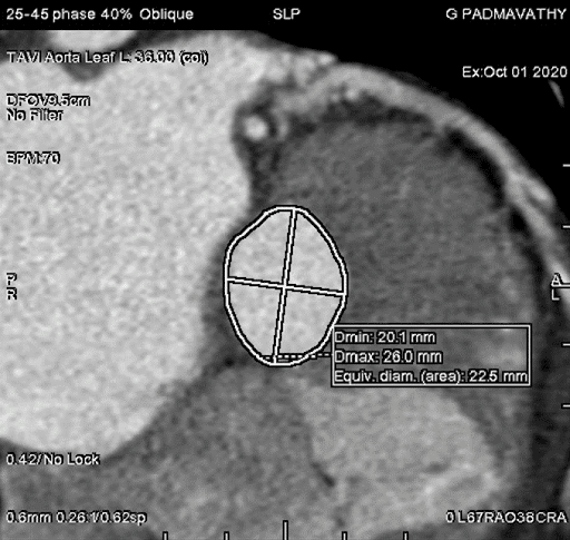
Before you undergo TAVR, your cardiologist will recommend various diagnostic tests to assess your overall health and risk factors. One of the most common tests that doctors prescribe is a CT angiogram.
balloon-expendable.jpg

TAVR is a new procedure that has been gaining popularity in recent years. It’s used for the treatment of aortic stenosis. It involves the use of a catheter to replace a damaged aortic valve with a biological tissue valve.


aortic-stenosis-1.jpg

Aortic valve stenosis is a serious condition that doesn’t just affect the aortic valve. It reduces or blocks the blood supply to the aorta. That, in turn, restricts blood flow to vital organs, including the brain, kidneys, and liver.


Echocardiogram-standard.jpeg

Grading Severity of Aortic Stenosis

Aortic valve stenosis is a serious disease that can lead to heart failure, strokes, and even death if left untreated. That makes timely diagnosis and treatment of the condition crucial. However, treatment of aortic stenosis depends on its severity.

Typically, the following parameters are used to determine the severity of aortic stenosis:

  • Pressure gradient – High gradient (HG; >/=40 mm Hg) or low gradient (LG; <40 mm Hg)
  • Blood flow – Normal flow (NF; SVi>35 ml/m2) or low flow (LF; SVi<35ml/m2)
  • Left ventricular ejection fraction (LVEF) – Preserved (>/=50%) or reduced (<50%)

Depending on the pressure gradient and blood flow parameters, aortic stenosis is graded as follows:

  • Normal flow-low gradient (NF-LG)
  • Normal flow-high gradient (NF-HG)
  • Low flow-high gradient (LF-HG)
  • Low flow-low gradient (LF-LG)

NF-HG is the most prevalent type of aortic stenosis and has well-established management protocols. Patients with NF-HG are also ideal candidates for aortic valve replacement. While LF-LG is fairly rare, it’s often associated with a poor prognosis.

Aortic-valve-stenosis 

Additionally, depending on progression, heart valve disease can be categorized into the following four stages:

  • Stage A (At risk) – Characterised by the presence of risk factors
  • Stage B (Progressive) – Mild or moderate valve disease with no noticeable symptoms
  • Stage C (Asymptomatic severe) – Severe valve damage with no noticeable symptoms
  • Stage D (Symptomatic severe) – Severe valve disease with noticeable symptoms

Doctors use a variety of diagnostic tests to evaluate the aforementioned parameters and determine the severity of aortic stenosis. If you experience symptoms like chest pain, heart murmur, or palpitation, it’s crucial to reach out to an experienced cardiologist and get the right treatment for aortic valve stenosis.

How Does One Diagnose Aortic Stenosis – ECG, ECHO, TEE, or CT aortogram?

Early diagnosis of aortic valve stenosis is crucial to prevent severe complications, such as arrhythmias, heart failure, stroke, and death. Also, it can help administer timely treatment, thus improving the patient’s prognosis and quality of life.

That’s why cardiologists use a series of tests to diagnose aortic valve stenosis and its underlying cause. When you visit the doctor, they’ll start by asking you about your symptoms and medical history. Also, they ask whether your family has a history of cardiovascular ailments. Next, they’ll use a stethoscope to detect the presence of the characteristic aortic stenosis murmur.

Additionally, your doctor will use one or more of the following tests for the complete diagnosis:

  • ECG (Electrocardiogram) – It’s one of the most preliminary tests that evaluate the heart’s electrical activity and helps doctors identify an irregular heartbeat and other abnormalities.
  • Echo (Echocardiogram) – It uses sound waves to generate images of the beating heart. It helps doctors examine how blood flows through each valve and determine the severity of aortic stenosis.
  • TEE (Transesophageal echocardiogram) – It’s a special type of echocardiogram in which an ultrasound probe is inserted into the esophagus and directed closer to the heart. It helps doctors take a closer look at the aortic valve and identify the underlying cause of aortic stenosis.
  • CT aortogram – It’s used to evaluate the blood supply to the upper body and identify conditions such as atherosclerosis.

Echocardiography (ultrasound) machine. doppler of aortic stenosis

Additionally, your doctor might recommend tests like cardiac catheterization and chest X-ray to get a complete picture of your cardiac health and plan the right course of treatment.

Book Online Consultaion


    Diagnosing Aortic Stenosis – Blog

    DOWNLOAD PDF

    Subscribe the Hearty Life Blogs


      Aotic-Valve.jpg

      Basics of the Heart

      The heart is a vital organ of the human body. It pumps blood throughout your body and keeps you alive. It comprises cardiac muscle tissue. That means the heart can contract and relax  to squeeze blood out of the heart and into your body. It supplies blood to all organs, including the brain, kidneys, liver, and more. 

      The heart has four chambers divided by two walls called septa. The upper chambers are called the atria, and the lower chambers are called the ventricles.

      The atria receive blood from the veins and pump it into the ventricles through openings called valves. The tricuspid valve separates the right atrium from the right ventricle, and the mitral valve separates the left atrium from the left ventricle. There’s also a pulmonary valve that sits between the right ventricle and the pulmonary artery.

      Then there’s the aortic valve located between the left ventricle and the aorta. It prevents blood from leaking back into the left ventricle during the contraction phase of the heart’s pumping cycle.

      When your heart beats, it squeezes blood out through an opening in each chamber called an aortic valve into either a large artery (aorta) or one of its branches, the carotid arteries, before heading back down to smaller arteries throughout your body. When the heart pumps blood to other organs, oxygen will be picked up by red blood cells as they pass through capillaries.

      What Is the Aortic Valve, and Why Is It Important?

      Aotic Valve

      The aortic valve is one of the four valves in the heart. It is a flap of tissue that keeps blood flowing in one direction. It is located in the middle of the heart, between the left ventricle and the aorta, the main artery that supplies blood throughout the body. The aortic valve is a semilunar valve, meaning the flaps of the valve are crescent-shaped.

      The aortic valve comprises three sections called leaflets or cusps. Each cusp is made of collagen. Some people are born with only two cusps in the aortic valve. In such cases, it is known as a bicuspid aortic valve.

      The aortic valve opens when the heart contracts or squeezes and closes when the heart relaxes. It lets oxygen-rich blood flow from the left ventricle into the aorta for circulation throughout your body. 

      The aortic valve is crucial because it ensures that blood keeps flowing in the right direction from the heart. It prevents the backflow of blood from the aorta into your left ventricle.

      If the aortic valve doesn’t work properly, it can cause serious problems. A leaky or stiffened valve won’t open and close properly, which means that some blood flows backward into the left ventricle. It puts extra pressure on the heart and can lead to heart failure or stroke if severe enough.

      Which Diseases Affect the Aortic Valve?

      The aortic valve is a crucial part of the heart that keeps blood flowing in the right direction throughout the body. The two primary diseases that affect the aortic valve are:

      • Aortic valve stenosis
      • Aortic valve regurgitation

      In aortic valve stenosis the cusps (or flaps) of the aortic valve become stiff and thick, thus narrowing the valve opening. That, in turn, restricts blood flow from the heart’s left ventricle to the aorta and the rest of the body.

      Aortic valve stenosis is usually caused by pre-existing conditions, such as congenital heart defects, aortic valve calcification, and rheumatic fever. Chronic ailments, such as hypertension, endocarditis, and diabetes, and treatments like radiation therapy to the chest can also increase the risk of aortic valve stenosis.

      In aortic valve regurgitation, the flaps don’t close properly, causing a backflow of blood into the left ventricle. As with aortic valve stenosis, this condition is also caused by pre-existing ailments that affect the heart.

      Common symptoms of aortic valve disease include heart murmur, chest pain, dizziness, fatigue, irregular heartbeat, and shortness of breath. Treatment for these conditions depends on the severity of the disease and its underlying cause. If left untreated, both aortic valve stenosis and aortic valve regurgitation can lead to heart failure and other complications.

      According to the CDC, nearly 2.5% of the US population suffers from heart valve diseases. These conditions are more common in older people, with 13% of people born before 1943 experiencing them. Also, 1% to 2% of the US population is affected by the bicuspid aortic valve, with the condition being more common in men.

      If you experience any of the aforementioned symptoms, reach out to an experienced cardiologist for proper diagnosis and treatment.

      Aortic Stenosis: What Is It?

      symptoms of Aortic Stenosis

      Aortic stenosis is a heart valve disease affecting the aortic valve that connects the heart’s left ventricle (lower chamber) to the aorta (main artery). The aortic valve is responsible for ensuring that blood flows in one direction throughout the body.

      In aortic valve stenosis, the aortic valve flaps (or cusps) become stiff or thick due to calcium buildup in the valve or other underlying causes. It causes the valve opening to become narrow and restricts blood flow to the aorta. That, in turn, reduces or blocks the blood supply to vital organs, such as the liver, kidney, brain, etc.

      Symptoms of aortic valve stenosis include:

      • A whooshing or swishing heart sound (known as aortic stenosis murmur)
      • Chest pain or tightness
      • Fatigue and dizziness (particularly after physical activity)
      • Shortness of breath
      • Palpitations

      Depending on the underlying cause, there are two types of aortic stenosis – congenital and acquired. Congenital aortic stenosis is present from birth due to a defect in the valve’s formation. Acquired stenosis develops after birth due to other heart-related ailments, such as hypertension, endocarditis, rheumatic fever, and aortic valve calcification.

      The risk of aortic stenosis increases in older people. Also, people who have received radiation therapy to the chest are more vulnerable to the condition. 

      Treatment for aortic valve stenosis depends on the underlying cause and the severity of the condition. Many patients need surgery to repair the valve. If left untreated, aortic stenosis can lead to heart failure, stroke, arrhythmias, and even death. Therefore, it’s crucial to diagnose aortic valve stenosis and treat the condition at the earliest.

      Book Online Consultaion


        Understanding Aortic Stenosis – Blog

        DOWNLOAD PDF

        Subscribe the Hearty Life Blogs


          Dr raghu logo


          Dr raghu logo


          +91 95424 75650

          Call us now if you are in a medical emergency need, we will reply swiftly and provide you with a medical aid.


          Call us now if you are in a medical emergency need, we will reply swiftly and provide you with a medical aid.


          Dr. Raghu | Heart Specialist in Hyderabad
          Yashoda Hospitals, Sardar Patel Rd, behind Hari Hara Kala Bhavan, Kummari Guda, Shivaji Nagar, Secunderabad, Telangana 500003

          Best Cardiologists in Hyderabad


          Copyright © 2023, Dr. Raghu. All rights reserved.

          [gtranslate]