CT-Coronary-Angiography-1.jpg

Doctors use various diagnostic tests to assess cardiac health and diagnose conditions like coronary artery disease and heart failure. While a coronary angiogram is commonly used to identify these conditions, your doctor can prescribe other tests like CT coronary angiography. 

In our previous blogs, you can read more about the use cases and risks of coronary angiography. In this article, we’ll delve deeper into CT coronary angiography and understand why it’s crucial for people with cardiac ailments.

What Is CT Coronary Angiography?

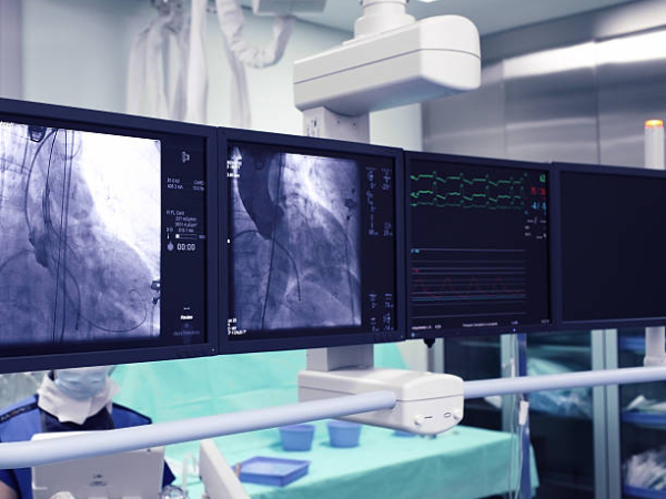
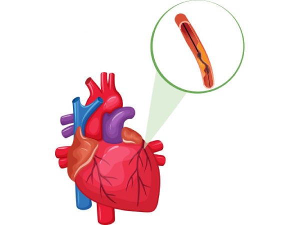
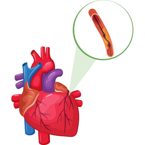
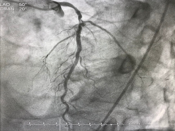
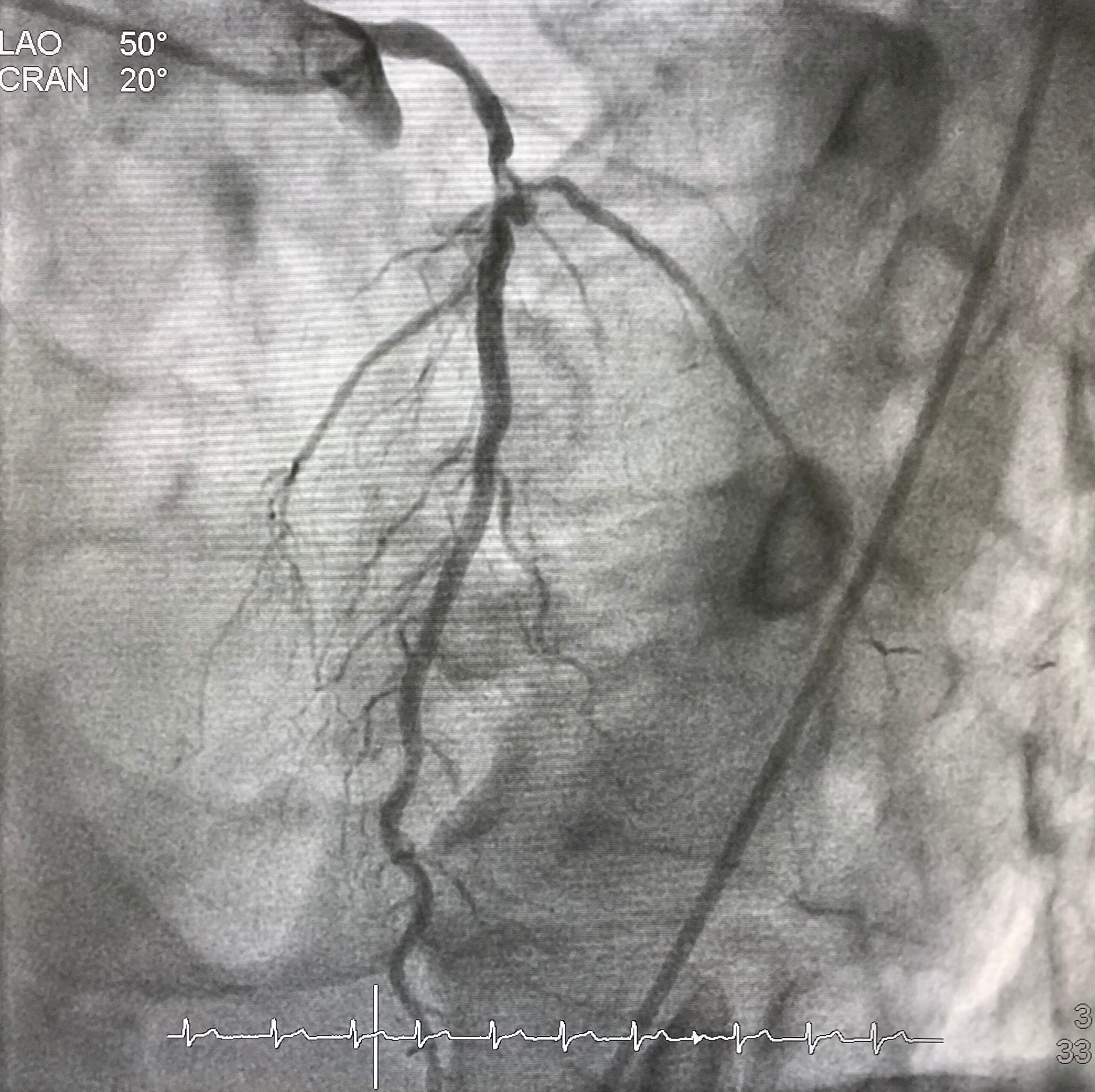
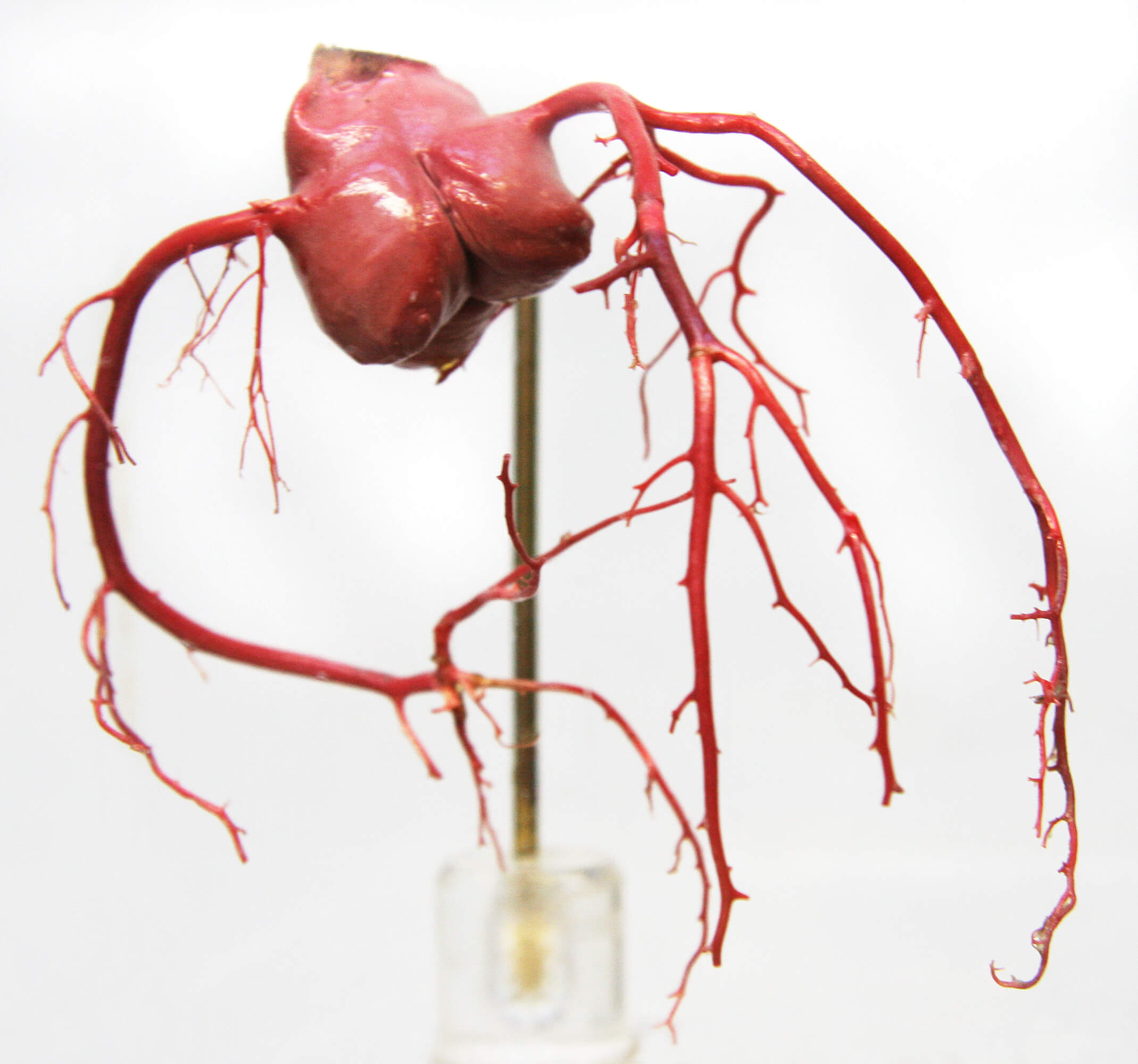
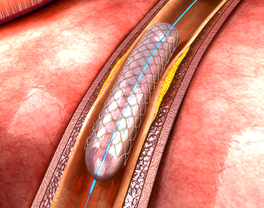
CT coronary angiography is a type of CT scan that uses special dyes to evaluate the heart’s blood vessels. It can detect blockages in the coronary arteries that supply blood to the heart. It can identify blockages that are too small for other methods, such as a regular angiogram or an electrocardiogram (ECG), to pick up.

CT coronary angiography

A CT coronary angiography scan aims to find any abnormal areas in your coronary arteries that could be causing stenosis (narrowing) or occlusion (blockage). It helps doctors identify underlying heart-related ailments and determine the right course of treatment.

How Does CT Coronary Angiography Work?

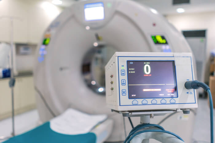
The test is usually done in a hospital or clinic. To begin with, the doctor or radiologist will start an intravenous (IV) line to administer the contrast dye. As the contrast flows through your veins, it will appear on the scan as bright white areas on a black background. It’ll help the doctor see your heart and coronary arteries. 

The test usually lasts about 30 minutes to one hour. The CT coronary angiogram procedure involves two scans: one with diastolic blood flow and another with systolic blood flow

When Is CT Coronary Angiography Used?

CT coronary angiography is used to diagnose a heart attack and evaluate blood flow in the heart. Also, doctors use it to detect blockages in arteries that supply blood to the heart muscle.

What Are the Risks of CT Coronary Angiography?

While CT coronary angiography is a non-invasive and safe procedure, it can cause the following side effects:

  • You may have an allergic reaction to the contrast dye.
  • Your body will be exposed to high levels of radiation, which can increase your risk of developing cancer.
  • You might have to pay out of pocket because the test isn’t always covered by insurance.
  • You might experience more anxiety and stress if the scans indicate blockages in your arteries.

What Should I Expect When Having CT Coronary Angiography?

If you are planning to get a CT coronary angiography, you should be prepared for the following:

  • The radiologist will ask you to change into a hospital gown.
  • The radiologist will give you an injection of a contrast dye.
  • You will lie on a table, and the table will move into the CT scanner.
  • The scan is painless and takes about 20 minutes.

Conclusion

CT coronary angiography is a painless and safe test that can help determine if you have blocked arteries. If you have chest pain, you should consider getting the test done as soon as possible because it may save your life.

Dr. C Raghu is a renowned cardiologist with decades of experience. He specializes in interventional cardiology and has treated thousands of patients with cardiac ailments. If you or anyone you know is experiencing symptoms like chest pain, breathlessness, palpitations, etc., reach out to Dr. Raghu today.

Book Online Consultaion


    CT Coronary Angiography – Blog

    DOWNLOAD PDF

    Subscribe the Hearty Life Blogs

      dr. raghu

      DR. RAGHU | CT Coronary Angiography in Hyderabad

      MD, DM, FESC, FACC, FSCAI

      Cardiology Coronary, Vascular and

      Structural Interventions


      Conditions & Diseases

      Coronary angioplasty

      Angioplasty

      Coronary angioplasty

      Aortic Stenosis

      Coronary angioplasty

      Atrial Fibrillation

      Coronary angioplasty

      Atrial Septal Defect


      View More Services


      New-Project-7.jpg

      Coronary angiography is a common diagnostic test used by doctors to identify conditions, such as coronary artery disease and aneurysms. In our previous blog, we discussed how the procedure is carried out and when it’s used. Click here to check it out.

      coronary angiography

      Angiography is a minimally invasive procedure, which makes it extremely safe. However, it can involve a few minor side effects. The benefits outweigh the risks for most patients. However, in some cases, coronary angiography can result in serious complications.

      In this article, we’ll delve deeper into the risks and side effects associated with coronary angiography.

      Common Side Effects of Coronary Angiography

      If you’re planning to visit the doctor for an angiography, you can expect one or more of the following side effects:

      • Bruising
      • Swelling
      • A buildup of blood (resulting in a bump)

      All these symptoms are localized to the area where the cut was made for inserting the catheter. Most patients experience a gradual improvement in these side effects without medical intervention. You can take painkillers to relieve discomfort after the procedure.

      Complications of Coronary Angiography

      If you’re lucky, you’ll come out of coronary angiography with minor bruising and swelling. However, some patients develop the following complications:

      • An infection near the cut :- It makes the area around the cut red, swollen, and tender.
      • An allergic reaction to the dye :- It usually results in an itchy rash.

      In both cases, proper use of medications can help control the side effects. For instance, antibiotics can be used in the case of an infection. Similarly, your doctor might prescribe antihistamines if you experience an allergic reaction.

      It’s worth mentioning that coronary angiography comes with a few potential serious complications. These include:

      • Kidney damage (from the dye)
      • Cardiac arrest or stroke
      • Internal bleeding (due to damaged blood vessels)
      • Anaphylaxis (due to severe allergic response to the dye)

      The good news is that these complications are extremely rare (affecting less than one in every 1000 patients). Also, kidney damage due to angiography is usually temporary. Moreover, internal bleeding can be contained with the help of catheter based approaches.

      Seeking Medical Help

      Complications from coronary angiography are rare. Nevertheless, it’s a good idea to consult your doctor if you notice anything unusual after the procedure. For instance, if the leg or arm where the cut was made looks pale or feels numb, it’s a cause for concern. Similarly, if you notice bleeding, redness, or a firm lump near the cut, it could indicate a potential infection. It’s always a good idea to watch out for these signs and consult your doctor for timely treatment.

      In Conclusion

      Coronary angiography is a safe and minimally invasive procedure. It can cause minor side effects, such as pain and swelling. However, in extreme cases, it can also lead to a heart attack or kidney damage. It’s crucial to talk to your doctor about the potential risks before going in for the procedure.

      Dr. C Raghu has more than two decades of experience in treating patients with different heart conditions. If you have queries or concerns about coronary angiography, feel free to reach out to Dr. Raghu today.

      Book Online Consultaion


        What Are the Risks of Coronary Angiography? – Blog

        DOWNLOAD PDF

        Subscribe the Hearty Life Blogs

          dr. raghu

          DR. RAGHU | Best Cardiologist in Hyderabad

          MD, DM, FESC, FACC, FSCAI

          Cardiology Coronary, Vascular and

          Structural Interventions


          Conditions & Diseases

          Coronary angioplasty

          Angioplasty

          Coronary angioplasty

          Aortic Stenosis

          Coronary angioplasty

          Atrial Fibrillation

          Coronary angioplasty

          Atrial Septal Defect


          View More Services


          Coronary-Bifurcation-Lesions.jpg

          Coronary arteries are the vessels which supply blood (oxygen and nutrients) to the heart; this supply may be disrupted due to buildup of cholesterol and fatty deposits, called plaques, on the inner walls of the arteries (atherosclerosis) causing narrowing of the vessel lumen.

          Coronary lesions branch out as they progress to supply various cardiac structures. A bifurcation lesion is a stenosis or abnormal narrowing of greater than 50% that occurs in a coronary artery at the origin of the side branch or adjacent to the origin of the side branch. Branching points a.k.a bifurcation points in the coronary arteries are prone to develop atherosclerosis as the shear stress is higher at the branch points.

          The figure shows how a bifurcation stenosis appears schematically as well as on angiography.

          bifurcation lesion


          Why bifurcation lesions are important?

          A vast majority of coronary arteries obstructions happen at the branching point. About 15-20% of patients of referrals for bypass surgery are because of the presence of coronary narrowing at bifurcation location. They are considered a tough lesion to treat by angioplasty and stent.

          How are bifurcation lesions diagnosed?

          Bifurcation lesions are identified on coronary angiography. Only lesions affecting side branch blood vessels more than 2.25 mm is considered important and called bifurcation lesions. The main vessel that gives branches is called main vessel and the branches that originate from it are called side branches. The point at the division of the main vessel and side branch is called a bifurcation point.

          How is a bifurcation lesion unique?

          Bifurcation lesions can involve the coronary artery either before or after the bifurcation point. In addition, side branches could be either diseased or free from disease. Based on the location and severity of the cholesterol plaque at the bifurcation point, sub types of bifurcation lesions have been identified.

          The challenge of performing angioplasty and stent for a bifurcation stenosis lies in the ability to preserve the main vessel and the side branch. Stents being metallic tubes risk occluding the side branch when placed across the side branch. This apprehension of losing the side branch, is one of the primary reasons for patients to be sent for bypass surgery instead of angioplasty and stent.

          Coronary-bifurication

          Can angioplasty and stent be performed for a bifurcation lesion?

          Angioplasty and stent can be eminently performed for a bifurcation stenosis. Over the last 2 decades numerous techniques in stent implantation have been developed to treat the bifurcation lesions. Some of these techniques include:

          • Provisional T stent technique
          • Tap technique
          • Double kiss crush technique
          • Culotte technique

          Advent of these techniques enabled experienced operators to perform angioplasty and stent successfully with chances of repeat narrowing seen in 3-6% patients at the site of stenting. Risk of losing a side branch after stenting is reduced to 1% once the new techniques are employed.

          Dr. C Raghu is an expert in bifurcation lesion angioplasty and the center where he has been trained, ICPS Paris France is considered the “Mecca” for bifurcation lesion angioplasty techniques development. 

          How does a bifurcation lesion angioplasty differ from a normal angioplasty?

          Bifurcation lesion angioplasty, involves utilization of novel stent techniques mentioned above. In addition, angioplasty operator experience is a major factor in determining  outcomes. Choosing the “right” stent that allows side branch access, use of specialized technologies such as fractional flow reserve (FFR), expertise in deploying 2 stents especially the process of “stent crush” and the handling of two balloons through a single catheter – kissing balloon are key points for the best results in a stent procedure.

          What are the chances of side branch occlusion in a bifurcation lesion angioplasty?

          Contemporary bifurcation angioplasty using Intravascular imaging employing contemporary stenting techniques in the hands of expert bifurcation angioplasty operators carries a 99% success rate with a chance of losing side branch in 1% and repeat narrowing at the end of 9 months being 3-6%. These results are comparable and if not superior to bypass surgery as angioplasty entails a short hospital stay and fast recovery.

          English Video

          Book Online Consultaion


            Coronary Bifurcation Lesions Blog

            DOWNLOAD PDF

            Subscribe the Hearty Life Blogs


              cci-coronary_angioplasty.jpg


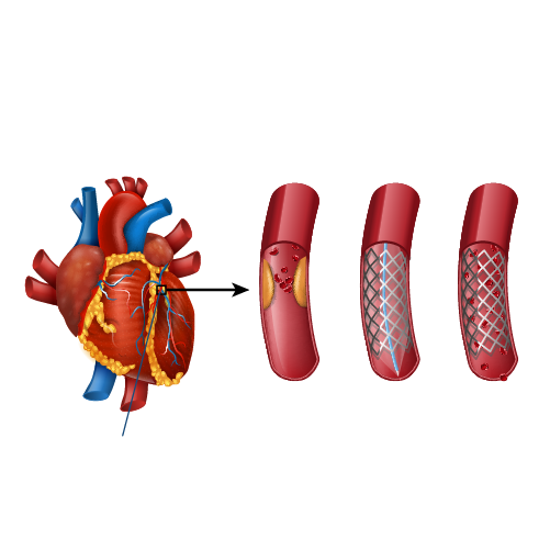
              Coronary arteries are the blood vessels that delivers oxygenated blood to the heart muscle. But, sometimes these blood vessels may get narrowed or blocked due to accumulation of cholesterol plaque. To open the blockage and restore the function of arteries, coronary angioplasty is performed.

              A coronary angioplasty involves the temporary insertion of a tiny balloon inside the artery to open the blockage. It is usually combined with the placement of a stent (a metal mesh tube) to widen the artery and prevent the further chance of narrowing.

              coronary angioplasty

              Why is coronary angioplasty done ?

              Coronary angioplasty is done to treat narrowing or blockage of the heart’s blood vessels:

              • during or after a heart attack.
              • Blood vessel narrowing leading to poor heart function.
              • For relief of chest pain (angina) due to reduced blood supply to heart.

              Risks of coronary angioplasty

              Usually, it is a safe procedure, but like all other procedures, it also carries certain risks like:

              Common Issues/Risks after angiography
              Bruising Common to have bruise at groin or arm Lasts for few weeks Not to be worried about
              Allergic reactions Usually related to contrast medicine used History of previous allergy – higher risk Present as rash and headache
              Bleeding at the site of entry (Hematoma) Usually subsides in a few days But in case swelling is increasing and painful contact your physician immediately


              Serious complications after coronary angiography Less than 1 in 1000 chance People with severe heart disease are at high risk Discuss with Cardiologist before procedure
              Heart attack A serious medical emergency where the heart’s blood supply is suddenly blocked
              Stroke A serious medical condition that occurs when the blood supply to the brain is interrupted
              Loss of blood supply to limb Damage to the artery in the arm or groin in which the catheter was inserted, with the blood supply to the limb possibly being affected
              Kidney injury Damage to the kidneys caused by the contrast dye
              Radiation injury Tissue damage caused by X-ray radiation if the procedure is prolonged

              What happens before the procedure?

              A physical examination and blood test are done to evaluate the overall health condition. The person needs to follow instructions provided before undergoing the procedure:

              1. Inform the doctor about:

              • Any allergies
              • The use of current medicines, including vitamin and mineral supplements
              • Any blood disorder
              • Any surgery you may had
              • The past and present medical condition

              2. Medicines:

              Tell the doctor if you are using blood thinners or any anti-diabetic medicines. The doctor will either advise you to stop or change the dose of the medication.

              3. Food and fluid restrictions:

              • Avoid heavy meals, such as meat, fried or fatty foods eight hours before the procedure. Take light foods such as toast and cereal.
              • Fast for six hours before the procedure.
              • Stay hydrated, drink plenty of fluid up to two hours before the procedure.

              In most of the cases, you will be discharged on the same day of the hospital, so ask someone to accompany you to the hospital.

              What happens during the procedure?

              Local anaesthesia would be administered to numb the area where the catheter would be inserted. Mostly, a catheter is inserted through the wrist, and sometimes through the groin. Under fluoroscopy (a type of X-ray), the catheter will be guided to the diseased artery. Then a special medicine (called contrast or dye) will be injected into the blood vessels supplying the heart. This will delineate the presence of obstruction to blood flow. 
              Then a wire is passed across the obstruction and a balloon is inflated at the site of obstruction to compress the obstructing plaque in the artery. The balloon expansion will compress the plaques into the wall of the blood vessel and improve the blood flow. Once the blood flow is improved, the catheter will be removed, and a stent would be placed to prevent the risk of further blockage. Finally, the catheter will be removed and the puncture site is sealed.

              What to expect after the procedure?

              The nurse will check the vitals of the patient. Depending on the condition, the doctor would advise you the hospital stay. You will be advised not to bend or cross the leg for a few days if the procedure is performed through the groin region. Additionally, some other tests like X-rays and an electrocardiogram (ECG) would be done to check the condition. Before discharge, the doctor will give you certain instructions regarding wound care, medications and lifestyle.

              Recovery after coronary angioplasty

              Recover quickly and keep your heart healthy by following these tips:

              • Quit smoking.
              • Limit the intake of alcohol.
              • Take the prescribed medicines.
              • Check the cholesterol levels regularly.
              • Maintain a healthy body weight.
              • Have a healthy and well-balanced diet.
              • Exercise regularly.

              Know your numbers

              It’s important to periodically monitor and maintain the following numbers to prevent future cardiac events

              • LDL cholesterol – less than 55 mg/dL
              • Blood pressure – less than 120/80 mm Hg
              • Blood sugar – HbA1C less than 7.0%

              Book Online Consultaion


                Download: Coronary Angioplasty Blog

                GET DOWNLOAD PDF NOW

                Subscribe the Hearty Life Blogs


                  Dr raghu logo


                  Dr raghu logo


                  Call us now if you are in a medical emergency need, we will reply swiftly and provide you with a medical aid.


                  For Appointment with Dr Raghu: +91 80085 36699
                  Dr Raghu’s Patient Coordinator: +91 72860 10203
                  Dr Raghu’s Office Experience: +91 90001 65962
                  Dr Raghu’s Social Media Communication Channel: +91 97046 51708

                  Dr. Raghu | Heart Specialist in Hyderabad
                  Yashoda Hospitals, Raj Bhavan Road, Somajiguda, 1st floor, Room No. 115, Hyderabad -500082, Telangana

                  Best Cardiologists in Hyderabad


                  © 2024, Dr. Raghu. All rights reserved. Design & Developed by AMSDigital.in

                  [gtranslate]
                  +91-9542475650