Resources | Dr Raghu - Page 6

cci-coronary_angioplasty.jpg


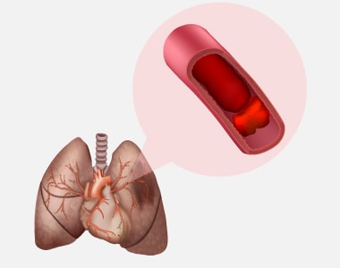
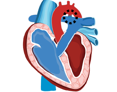
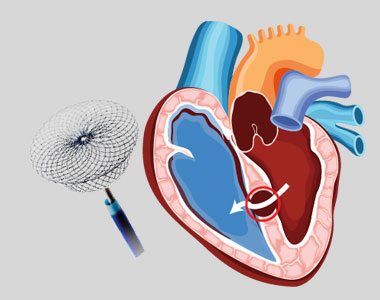
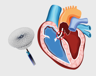
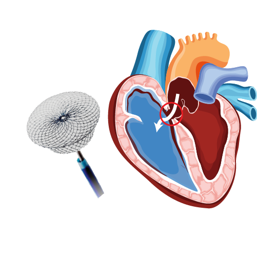
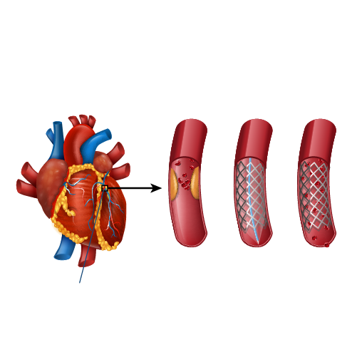
Coronary arteries are the blood vessels that delivers oxygenated blood to the heart muscle. But, sometimes these blood vessels may get narrowed or blocked due to accumulation of cholesterol plaque. To open the blockage and restore the function of arteries, coronary angioplasty is performed.

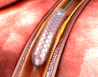
A coronary angioplasty involves the temporary insertion of a tiny balloon inside the artery to open the blockage. It is usually combined with the placement of a stent (a metal mesh tube) to widen the artery and prevent the further chance of narrowing.

coronary angioplasty

Why is coronary angioplasty done ?

Coronary angioplasty is done to treat narrowing or blockage of the heart’s blood vessels:

  • during or after a heart attack.
  • Blood vessel narrowing leading to poor heart function.
  • For relief of chest pain (angina) due to reduced blood supply to heart.

Risks of coronary angioplasty

Usually, it is a safe procedure, but like all other procedures, it also carries certain risks like:

Common Issues/Risks after angiography
Bruising Common to have bruise at groin or arm Lasts for few weeks Not to be worried about
Allergic reactions Usually related to contrast medicine used History of previous allergy – higher risk Present as rash and headache
Bleeding at the site of entry (Hematoma) Usually subsides in a few days But in case swelling is increasing and painful contact your physician immediately

Serious complications after coronary angiography Less than 1 in 1000 chance People with severe heart disease are at high risk Discuss with Cardiologist before procedure
Heart attack A serious medical emergency where the heart’s blood supply is suddenly blocked
Stroke A serious medical condition that occurs when the blood supply to the brain is interrupted
Loss of blood supply to limb Damage to the artery in the arm or groin in which the catheter was inserted, with the blood supply to the limb possibly being affected
Kidney injury Damage to the kidneys caused by the contrast dye
Radiation injury Tissue damage caused by X-ray radiation if the procedure is prolonged

What happens before the procedure?

A physical examination and blood test are done to evaluate the overall health condition. The person needs to follow instructions provided before undergoing the procedure:

1. Inform the doctor about:

  • Any allergies
  • The use of current medicines, including vitamin and mineral supplements
  • Any blood disorder
  • Any surgery you may had
  • The past and present medical condition

2. Medicines:

Tell the doctor if you are using blood thinners or any anti-diabetic medicines. The doctor will either advise you to stop or change the dose of the medication.

3. Food and fluid restrictions:

  • Avoid heavy meals, such as meat, fried or fatty foods eight hours before the procedure. Take light foods such as toast and cereal.
  • Fast for six hours before the procedure.
  • Stay hydrated, drink plenty of fluid up to two hours before the procedure.

In most of the cases, you will be discharged on the same day of the hospital, so ask someone to accompany you to the hospital.

What happens during the procedure?

Local anaesthesia would be administered to numb the area where the catheter would be inserted. Mostly, a catheter is inserted through the wrist, and sometimes through the groin. Under fluoroscopy (a type of X-ray), the catheter will be guided to the diseased artery. Then a special medicine (called contrast or dye) will be injected into the blood vessels supplying the heart. This will delineate the presence of obstruction to blood flow. 
Then a wire is passed across the obstruction and a balloon is inflated at the site of obstruction to compress the obstructing plaque in the artery. The balloon expansion will compress the plaques into the wall of the blood vessel and improve the blood flow. Once the blood flow is improved, the catheter will be removed, and a stent would be placed to prevent the risk of further blockage. Finally, the catheter will be removed and the puncture site is sealed.

What to expect after the procedure?

The nurse will check the vitals of the patient. Depending on the condition, the doctor would advise you the hospital stay. You will be advised not to bend or cross the leg for a few days if the procedure is performed through the groin region. Additionally, some other tests like X-rays and an electrocardiogram (ECG) would be done to check the condition. Before discharge, the doctor will give you certain instructions regarding wound care, medications and lifestyle.

Recovery after coronary angioplasty

Recover quickly and keep your heart healthy by following these tips:

  • Quit smoking.
  • Limit the intake of alcohol.
  • Take the prescribed medicines.
  • Check the cholesterol levels regularly.
  • Maintain a healthy body weight.
  • Have a healthy and well-balanced diet.
  • Exercise regularly.

Know your numbers

It’s important to periodically monitor and maintain the following numbers to prevent future cardiac events

  • LDL cholesterol – less than 55 mg/dL
  • Blood pressure – less than 120/80 mm Hg
  • Blood sugar – HbA1C less than 7.0%

Book Online Consultaion


    Download: Coronary Angioplasty Blog

    GET DOWNLOAD PDF NOW

    Subscribe the Hearty Life Blogs


      disease-condtions-pulmonary_embolism.jpg

      Pulmonary embolism is a blockage in one of the pulmonary arteries of the lungs. The most common reason for pulmonary embolism (PE) is a blood clot (thrombus) that travels to the lungs from other parts of the body (usually a leg).


      disease-condtions-deep_vein_thrombosis.png

      When a blood clot (thrombus) is formed in one or more of the deep veins of your body (usually in the legs), it is known as deep vein thrombosis (DVT). Sometimes this clot becomes detached from the inner walls of the blood vessel and travels to another part inside the body. DVT may develop due to an underlying medical condition particularly the one which affects the clotting of the blood.

      Causes and risk factors:

      It can be caused by the factors which prevent the normal circulation of the blood and it’s clotting, such as long periods of rest (during a particular medical condition), injury to the vein, surgery and certain medications.

      The risk factors are as follows:

      • Inherited blood clotting disorder
      • Prolonged bed rest
      • Surgery
      • Pregnancy
      • Contraceptives or hormone replacement therapy
      • Obesity
      • Smoking
      • Cancer
      • Heart failure
      • Inflammatory bowel disease
      • Age (usually older than 60 years but can also occur at any age)

      Complications:

      The following are the complications which may arise due to DVT:

      Pulmonary embolism: In Pulmonary embolism, a thrombus present in another part of your body usually a leg, travels to the blood vessel of the lung and blocks the blood circulation, leading to a life-threatening condition.

      Post-phlebetic syndrome (post thrombotic syndrome): set of signs and symptoms seen after a blood clot has formed.

      Symptoms:

      The person may be asymptomatic during the formation of the thrombus, however, during post thrombotic period when it has caused damage to the veins, the blood flow to the affected body part may be compromised. The following symptoms are observed:

      • Persistent swelling of the leg or arm (edema)
      • Leg pain or tenderness while standing
      • Skin discoloration in the affected arm or leg
      • Veins in the arm or leg appearing larger than normal

      Sometimes the development of a serious health complication such as pulmonary embolism is responsible for the symptoms in the DVT patients which are as follows:

      • Shortness of breath
      • Chest pain
      • Cough (blood may be coughed out as well)
      • Back pain
      • Profuse sweating
      • Lightheadedness
      • Blue nails or lips

      These symptoms appear suddenly, and an immediate medical attention is required.

      Diagnosis:

      The diagnosis is initiated by a thorough clinical examination and involves review of the patient’s medical history. Based upon these findings, the physician will categorize the patient into low or high risk DVT. Further tests may be considered to rule out other health problems and to confirm the diagnosis, which are as follows:

      • Blood tests: These are considered to evaluate any blood disorders that increase risk of DVT.
      • D- dimer test: In this test, the levels of a protein called D-dimer is assessed. In patients with DVT, high levels of this protein can be found in the blood.
      • Duplex ultrasonography: It is a non-invasive evaluation of the blood flow in the arteries and the veins of a patient. But this test is not preferred for a thrombus located very deep inside the body such as pelvis.
      • Venography: It is a specialized type of an x-ray procedure wherein a dye is injected into a vein of the foot, and the blood clot if present is detected.
      • Magnetic resonance imaging (MRI): It is performed in patients for whom ultrasound examination is inappropriate or unfeasible. It can even detect a thrombus present in the patient’s pelvis and the thigh.

      Prevention:

      A recent World Thrombosis Day Ipsos survey showed that there is less awareness of DVT in countries around the world. Thus, the first step in prevention is making the public aware of DVT. The preventive measures are different depending on the person’s condition who are at risk of developing DVT:

      During pregnancy

      In pregnant women, certain medication may be prescribed if she has risk factors such as likelihood of having a cesarean birth, bed rest, history of DVT, and inherited thrombophilia.

      During travel or in longer hours of sitting posture:

      • Drink lots of fluids.
      • Wear loose fitting clothes.
      • Walk and stretch at regular intervals.
      • Use special stockings to improve blood flow and to prevent blood clots but consult your health care provider before using it.

      Treatment:

      Treatment for DVT includes certain medicines and therapies, which are as follows:

      Medications:

      • Anticoagulants: They prevent further formation of blood clots but don’t dissolve the already existing ones. Most common anticoagulants used are heparin and warfarin. Warfarin is not recommended during pregnancy.
      • Thrombin inhibitors: These are recommended for people who can’t take heparin.
      • Thrombolytic medicines: These are recommended only during life threatening condition.

      Compression stockings:

      It may be recommended to prevent swelling and to avoid post-thrombotic symptoms in the patients.

      Surgical treatment:

      Surgical treatment may be performed when medications fail to dissolve the clots.

      Book Online Consultaion


        Deep Vein Thrombosis Blog

        DOWNLOAD PDF

        Subscribe the Hearty Life Blogs


          disease-condtions-thromboembolism.jpg

          Thromboembolism refers to a condition where a blood clot formed in the blood vessel unplugs itself and flows in the blood stream to block another blood vessel leading to obstruction of blood flow. Although clotting is a normal bodily function, if a clot occurs where it is not required, it may lead to serious problems.

          Types of Thromboembolism:

          The type of thromboembolism differs depending on the area in which the clot may appear. It can obstruct the blood flow in the veins, arteries, brain, gastrointestinal tract or kidneys.

          Thromboembolism can be further classified as below:

          Venous Thromboembolism:

          A venous thromboembolism is a thrombus (blood clot) formed in a vein. The blood flow in the veins is slow when compared to arteries which can increase the likelihood of the blood clots forming in these vessels.

          It can be categorized into three different types

          • Superficial Vein Thrombosis
            The clot may occur in a superficial vein, close to the surface of the body. Usually, it is not serious until it enters the deep veins.
          • Deep Vein Thrombosis (DVT)
            Deep vein thrombosis occurs within the deep veins, and mostly occurs within the legs.
          • Pulmonary embolism (PE)
            Pulmonary embolism is a medical emergency in which the thrombus blocks the blood vessel in the lungs.

          Renal Vein Thrombosis (RVT)

          A renal vein thrombosis is a thrombosis that occurs in the veins that drain blood away from the kidneys. These clots reduce the ability of the kidneys to clean and filter the blood.

          Arterial Thrombosis (atherothrombosis)

          Arterial thrombosis is much less common than venous thrombosis. It can have similar risks. Usually arterial thrombosis may lead to necrosis of the tissue.

          A thromboembolism in the coronary artery can cause a heart attack. If blood supply to the brain is disrupted, the patient may suffer a stroke.

          Symptoms:

          Signs and symptoms of venous thromboembolism include the following

          DVT

          • Pain in arm or leg, usually in the thigh or calf
          • Swollen leg or arm
          • Reddening or warming of the skin
          • Red streaks on the skin

          PE

          • Shortness of breath
          • Chest pain under your rib cage that can get worse when you take a deep breath
          • Rapid heart rate
          • Feeling lightheaded or passing out
          • Accentuated second heart sound

          Arterial Thrombosis

          • Muscle pain or spasm in the affected area
          • Numbness and tingling in the arm or leg
          • Pale color of the arm or leg (pallor)
          • Weakness of an arm or leg

          If the condition is severe, it may lead to blister formation and shedding of the skin, leading to tissue necrosis.

          Symptoms of a clot in an organ vary with the organ involved but may include:

          • Pain in the part of the body that is involved
          • Temporary decrease in organ function

          Causes:

          The blood clots can occur due to injury to a vein, consequence of a surgery, use of certain medications and lack of movement of the limbs.

          In the case of PE, the blood clot may block the blood vessels of the lungs. The affected portion of the lung may die due to loss of blood supply, the condition is called as pulmonary infraction, making it difficult to provide oxygen to the rest of the body.

          Occasionally, the blood vessels can be blocked by substances other than blood like collagen, part of a tumour, air bubbles etc.

          Risk factors:

          The common risk factors include

          • Inheriting a blood-clotting disorder
          • Prolonged immobility which may be due to bed rest or long trips
          • Injury or surgery
          • Pregnancy
          • Birth control pills or hormone replacement therapy
          • Being overweight or obese
          • Smoking
          • Cancer
          • Heart failure
          • Inflammatory bowel disease
          • Family history of deep vein thrombosis or pulmonary embolism

          Diagnosis:

          The diagnosis of thromboembolism includes the following tests which could be considered depending on the type:

          D-dimer: The blood sample is tested for the presence of D-dimer which is a marker for the presence of blood clots.

          Duplex Ultrasound: This is an imaging test in which the presence of clots is identified using ultrasound waves.

          Pulse oximetry: In this test, a sensor attached on the end of the finger of the patient helps to measure the level of oxygen in the blood.

          Arterial blood gas: The blood drawn from the artery is checked for oxygen levels present in it.

          Chest X-ray: This test may not be useful in finding the presence of clots, but can help to rule out a clot.

          Other diagnostic tests may include ELISA, pulmonary angiography, venography, echocardiography, helical computed tomography of pulmonary vessels etc.

          Treatment:

          Treatment of thromboembolism includes:

          Blood thinners: These are anticoagulant drugs which prevent formation of new clots while the body works to break up the previous clots present. They include heparin, low-molecular-weight heparin, apixaban, edoxaban, rivaroxaban, and warfarin.

          Clot-busting drugs: These are intravenous injections to dissolve the clots in the case of life-threatening situations. They include drugs belonging to the class of tissue plasminogen activators.

          Surgical and other procedures:

          • Removal of clot: Surgery is considered only if the condition is life threatening. The clot is removed using a catheter threaded through the blood vessel.
          • Vein filter: A catheter can also be used to position a filter in the inferior vena cava which helps to stop the clots from being carried into the lungs.

          Prevention:

          If the patient is at a risk of the condition, the following preventive measures may be considered to reduce the occurrence:

          • Use of blood thinners
          • Use of compression stockings
          • Use of intermittent pneumatic compression devices that automatically squeeze the legs to keep blood flowing

          Other preventive measures include

          • Regular exercise
          • Maintaining healthy weight
          • Cessation of smoking

          Consideration during travel include

          • Walk every 1 to 2 hours
          • Stretch legs and move around on the seat
          • Drink lots of fluids
          • Avoid smoking and consumption of alcohol before journey

          Book Online Consultaion


            Thromboembolism Blog

            DOWNLOAD PDF

            Subscribe the Hearty Life Blogs


              disease-condtions-varicose_veins.jpg

              Varicose veins are the twisted, swollen and bulging veins that appear in blue, red and skin color, just beneath the surface of the skin. They occur mostly in the legs and feet, causing aching pain and discomfort in the legs.


              disease-condtions-pda.png

              Patent ductus arteriosus (PDA) is a congenital heart condition in which there is an opening between the pulmonary artery and the aorta. The ductus arteriosus is a small connection in the fetal heart helps oxygen rich blood to by-pass the immature baby’s lungs and flow into the body.


              disease-condtions-device_closure_for_vsd.jpg

              The heart consists of four chambers, of which upper two chambers are called as atria and lower two chambers are called as ventricles. Right and left chambers are separated by a wall of muscle called a septum. The septum separating the atria is called inter atrial septum and the once between the two ventricles in inter ventricular septum.

              Right two chambers – right atrium and right ventricle pump the deoxygenated blood collected from various parts of the body to the lungs. Blood gets saturated with oxygen in the lungs and returns back to the left side of the heart.

              Ventricular septal defects (VSD) is a common type of congenital heart defect, which is characterized by an abnormal opening or a hole in the interventricular septum, the dividing wall between right and left ventricles. The oxygen-rich blood from the left ventricle will enter into the right ventricle through the opening, thereby getting mixed with deoxygenated blood which then enters into lungs. This will force the heart and lungs to work harder.

              Causes:

              The exact cause of VSD is unknown. During fetal heart developmental stage, the heart develops from a large tube which eventually divides into chambers and walls. Any abnormality in this process will lead to the formation of a defect in the septum. If the defect is in the interventricular septum, then it is said to be ventricular septal defect. There may be one or more VSDs.

              ventricular septal defects

              Types:

              Based on the location and development of VSD, it is classified into following types:

              • Conoventricular Ventricular Septal Defect: It occurs just below the pulmonary and aortic valves.
              • Perimembranous Ventricular Septal Defect: It occurs in the upper part of the ventricular septum.
              • Inlet Ventricular Septal Defect: It occurs adjacent to the tricuspid and mitral valves. This type of defect might be associated with atrial septal defect.
              • Muscular Ventricular Septal Defect: It is the most common type of VSD, which occurs in the lower muscular part of the interventricular septum.

              Signs and symptoms:

              Small defects in the septum do not show any symptoms because it closes on its own gradually during childhood.

              Large defects show signs and symptoms usually after birth within a few weeks or months.

              The first sign of VSD is heart murmur, which is a whooshing sound that can be heard using a stethoscope. The other symptoms include:

              • Fatigue (tiredness)
              • Arrhythmias (abnormal heart rhythm)
              • Fast breathing or breathlessness
              • Poor feeding
              • Poor weight gain
              • Pale skin
              • Enlarged liver

              Risk factors:

              Ventricular septal defects mostly occur due to defective genes and chromosomes, that may be hereditary.

              Environmental factors during pregnancy may also play a role in development of VSD in the fetus.

              Complications:

              Large or medium septal defects if left untreated, may lead to life threatening complications such as:

              • Heart failure, as heart need to work harder to pump enough blood to the body.
              • Pulmonary hypertension (increased blood flow to the lungs results in increased blood pressure in the lung arteries).
              • Endocarditis (infection in the endocardium of heart).
              • Other heart problems such as abnormal heart rhythms and valve problems.

              ventricular septal defects

              Diagnosis:

              If heart murmurs are detected during the physical examination, the patient may be advised further testing to conform the diagnosis.

              • Echocardiogram: This test is the primary tool for the diagnosis of VSD, as it can be used to determine the size, location and severity of the ventricular septal defect. Sound waves are used to produce the detailed images of the heart.
              • Electrocardiogram (ECG): This test records the electrical activity of the heart and helps to identify the abnormal heart rhythms and defects in the septum.
              • Chest X-ray: X-rays are used to view the images of heart and lungs. In VSD, this test helps to determine the enlarged heart and extra fluid in the lungs.
              • Cardiac catheterization: The test involves inserting a thin, flexible tube into the blood vessel at the groin or an arm, which is guided to the heart, to identify any congenital heart defects and to determine the function of the heart chambers and its valves.
              • Pulse oximetry: Oxygen levels in the blood can be measured using a small clip which is placed on the fingertip.

              Treatment:

              Usually treatment is not needed for small VSDs, as they close on their own gradually after birth. 

              Babies with larger VSD need surgery to prevent any further complications. 

              The treatment for ventricular septal defects include:

              • Medications such as diuretics like furosemide are used to reduce the amount of fluid in the blood that is pumped to the lungs and in the circulation. Beta blockers like metoprolol, propranolol and digoxin are used to maintain the regular heartbeat.
              • Surgical procedures include surgical repair, catheter procedure and hybrid procedure.
                Surgical repair involves open heart surgery, in which the doctor uses a patch or stitches to close the hole, performed under general anaesthesia.
                In catheter procedure, a catheter is inserted into a blood vessel and then passed to the heart. The defect is closed using a specially sized mesh device.
                Hybrid procedure uses both surgical repair and catheterization procedures to close the hole, with the help of a heart-lung machine and a catheter placed through an incision.

              Prevention:

              Ventricular septal defects cannot be prevented, but following certain measures during pregnancy may be helpful to prevent the risk of VSD. The measures include:

              • Prenatal care before pregnancy: Consult a physician before planning for pregnancy and inform him about the family history of any congenital defects and the medications using currently, so that he may give suggestions or recommend some lifestyle modifications to avoid the risk of heart defects.
              • Healthy diet: Having a balanced diet including vitamins and folic acid during pregnancy will help in giving birth to a healthy child without any heart defects.
              • Regular exercise under the supervision of a gynecologist is necessary during pregnancy.
              • Avoid alcohol, tobacco and harmful drug use : during pregnancy to prevent the risk of VSD.
              • Get vaccinated: Check whether you are vaccinated for vaccine-preventable infections before getting pregnant.
              • Control diabetes: Monitor your sugar levels regularly to prevent the risk of heart defects.
              English Video

              Book Online Consultaion


                Ventricular Septal Defects Blog

                DOWNLOAD PDF NOW

                Subscribe the Hearty Life Blogs


                  disease-condtions-device_closure_for_asd.jpg

                  What is an Atrial Septal Defect or ASD? 

                  Heart consists of four chambers, of which upper two chambers are called as atria and lower two chambers are called as ventricles. Atrial septal defect (ASD) is a type of congenital heart defect, in which there is an abnormal opening or a hole in interatrial septum (dividing wall between two atria). This opening allows the passage of pulmonary venous blood from left atrium to right atrium, causing mixing of oxygenated and deoxygenated blood in right atrium and increasing the flow of blood to lungs. The increased blood flow may increase the workload of the lungs, and eventually cause damage to heart and lungs.

                  What causes an ASD?

                  The exact cause of ASD remains unclear. However, it is believed that during fetal developmental stages, a hole is present in the interatrial septum, which gradually closes before birth or during infancy. If the hole persists, it is called an atrial septal defect.

                  Types of Atrial Septal Defect or ASD?

                  Based on the location and development of ASD, it is classified into four types:

                  • Ostium secundum ASD: It occurs in the middle part of the interatrial septum. This is the most common type of ASD and accounts for 75% of all atrial septal defects. This type of ASD is commonly detected in adults in their third and fourth decades of life. Some can be detected in children when an abnormal heart sound is detected at the time of a routine health check or vaccination visit. 
                  • Ostium primum ASD (20%): It occurs in the lower part of the interatrial septum, adjacent to atrioventricular (AV) valves. It usually occurs as a part of other congenital heart defects. This defect is usually detected in early life as this is associated with many complications.
                  • Sinus venosis ASD (4%): It occurs in the upper part of the interatrial septum, near the veins that drain into the right and left atrium. This is usually identified in third and fourth decade adults. 
                  • Coronary sinus ASD (<1%): It occurs in the interatrial septum between the coronary sinus and the left atrium. This is very uncommon and patients are asymptomatic.

                  Signs and symptoms

                  Usually after birth, babies who have ASD may not show any associated signs and symptoms. But, symptoms may appear during adulthood around the age of 30 years. Most of them don’t have any symptoms even after many years.

                  Some of the common symptoms associated with ASD are:

                  • Heart murmur, a swishing sound that can be heard through a stethoscope
                  • Heart palpitations
                  • Arrhythmias (abnormal heart rhythms)
                  • Fatigue (feeling tired mainly after exercise)
                  • Shortness of breath
                  • Swelling of legs, feet or abdomen
                  • Stroke
                  • Lung infections such as pneumonia

                  Risk factors:

                  Genetics and environmental factors usually play a role in congenital heart defects. Some conditions during pregnancy may confer high risk for heart defects, 

                  Complications of an untreated ASD:

                  • Right-sided heart failure
                  • Arrhythmias
                  • Increased risk of a stroke
                  • Shortened life span
                  • Pulmonary hypertension (increased blood flow to the lungs results in increased blood pressure in the lung arteries)
                  • Eisenmenger syndrome (pulmonary hypertension causing permanent lung damage)

                  Diagnosis:

                  Most ASDs are diagnosed incidentally during regular heath check-ups. If a heart murmur is heard during auscultation, you may be advised to undergo further tests to confirm the diagnosis of ASD: 

                  • Echocardiogram: It is a specific diagnostic test for ASD, and provides a detailed image of the heart and blood flow through its chambers. The echocardiogram may show the blood flow through the interatrial septum and the size of defect in the septum.
                  • Chest X-ray: It helps to identify enlarged heart and lung changes.
                  • Electrocardiogram (ECG): It involves recording the electrical activity of the heart, to help identify arrhythmias.
                  • Cardiac catheterization: A thin, flexible tube called catheter is inserted into the blood vessel at the groin or arm and is guided to the heart. This test is helpful to determine the function of heart and its valves, and to measure the blood pressure in the lungs.
                  • Magnetic resonance imaging (MRI): It uses magnetic and radio waves to create three dimensional images of the heart and other organs. This test is recommended if the ASD is not clearly diagnosed with echocardiogram.
                  • Computerized tomography (CT) scan: It uses a series of X-rays to create detailed images of heart; mainly used if echocardiogram doesn’t help to diagnose ASD clearly.

                  Treatment:

                  Treatment of ASD depends on the age at diagnosis and the size, location and severity of the defect. Small ASDs might not need any treatment because it closes on its own. The doctor may recommend surgery if the atrial septal defect is large, even with fewer symptoms to prevent problems later in life. Treatment of ASD includes medications and surgery.

                  • Medications usually do not help in closing the hole, but reduces the signs and symptoms associated with ASD and risks of complications after surgery. Drugs such as beta blockers (to maintain regular heartbeat) and anticoagulants (to prevent the formation of blood clots) are used.
                  • Surgery includes cardiac catheterization and open-heart surgery.

                  Device closure of ASD

                  In cardiac catheterization, the doctor closes the hole in the septum with a mesh patch or a plug in the defect place using a catheter. The heart tissue then grows around the mesh slowly, sealing the hole permanently. This procedure is mainly performed to repair only the secundum type of atrial septal defects. Defects not amenable to closure with device may need open-heart surgery.

                  In open-heart surgery, the defect may be closed with stitches or a special patch. This procedure is done under general anesthesia, mainly for repairing primum, sinus venosus and coronary sinus atrial septal defects.

                  Prevention of ASD:

                  Atrial septal defects cannot be prevented, but following certain measures during pregnancy might be beneficial to prevent the risk of ASD, such as:

                  • Immunity test for rubella: If the person is not immune to rubella, it is necessary to get vaccinated.
                  • Monitoring current health conditions and medications use: Pre-existing health conditions and usage of any medications should be carefully monitored during pregnancy to prevent the risk of ASD.
                  • Reviewing family medical history: If a person has a family history of congenital defects, it is advised to visit a genetic counsellor before becoming pregnant to know the risks of atrial septal defects.
                  English Video


                  Our Specialities


                  • 24 hours emergency services
                  24 hours emergency services
                  • Clinics- weekly basis/monthly basis/ Yearly basis
                  Clinics- weekly basis/monthly basis/ Yearly basis
                  • Prevention of cardiovascular diseases
                  Prevention of cardiovascular diseases
                  • Diagnosis
                  Diagnosis

                  BOOK AN APPOINTMENT

                    dr. raghu

                    DR. RAGHU

                    MD; DM; FESC; FACC; FSCAI
                    Cardiology Coronary, Vascular and
                    Structural Interventions


                    Conditions & Diseases

                    Coronary angioplasty

                    Angioplasty

                    Coronary angioplasty

                    Aortic Stenosis

                    Coronary angioplasty

                    Atrial Fibrillation

                    Coronary angioplasty

                    Atrial Septal Defect


                    View More Services

                    disease-condtions-coronary_artery_disease.jpg

                    Coronary artery disease (CAD) is the most common type of heart disease, in which the coronary arteries ( blood vessels that supplies oxygen and nutrients to the heart muscle) become rigid.


                    Acute myocardial infraction is a sudden obstruction in the blood flow to the heart muscle. It is an emergency condition in which the plaque formed due to the deposition of fat, cholesterol, calcium, or cellular waste products is ruptured leading to the formation of thrombus. This leads to the obstruction of the coronary vessels resulting in an acute reduction of blood supply.

                    Signs and Symptoms:

                    The symptoms of Acute myocardial infraction include:

                    • Chest Pain- Uninterrupted and intense chest pain often radiates to the neck, shoulder, jaw, and down to the left arm. The chest pain is usually experienced as pressure, squeezing, aching, or burning sensation, and can also be presented as a feeling of indigestion or fullness of gas.

                    The important signs of the patient to be considered include the following:

                    • Increase in the heart rate
                    • Irregular pulse
                    • Elevated blood pressure
                    • Coughing and wheezing
                    • Nausea and vomiting
                    • Shortening of breath
                    • Distended neck veins
                    • Production of frothy sputum
                    • Excessive sweating, light headedness and palpitations.
                    • Loss of consciousness due to inadequate blood flow to the brain

                    Causes:

                    Coronary artery blockage is the primary cause of acute myocardial infarction. It is due to the accumulation of cholesterol in the arteries. The increase in LDL levels (bad cholesterol) in the body leads to plaque formation in the arteries. Saturated and trans fats are other types of fats which also lead to the formation of plaque in the arteries and obstruct the blood flow. Certain dairy products including butter and cheese, meat, beef, and processed foods are the main sources of saturated and trans fats.

                    During a heart attack, the plaque gets ruptured and spills cholesterol into the bloodstream. This leads to the formation of blood clots, when are big enough can block the artery at the site of rupture and deprives the heart muscle from obtaining enough oxygen and nutrients. There can be a partial block of the artery or a complete block of the artery. A complete block refers to an ST level elevation Myocardial Infraction (STEMI) and a partial block refers to a non-ST elevation Myocardial Infraction (NSTEMI).

                    Risk Factors:

                    Some of underlying risk factors of the acute myocardial infraction are modifiable.

                    • Sex
                    • Age
                    • Family History
                    • Male Pattern Baldness
                    • Smoking
                    • Diabetes Mellitus
                    • Hypertension
                    • Obesity
                    • Lack of exercise
                    • Poor oral hygiene
                    • Presence of peripheral vascular disease
                    • Elevated levels of homocysteine

                    Other factors include

                    • Trauma
                    • Drug use (Cocaine)
                    • Vasculitis
                    • Coronary artery anomalies
                    • Coronary artery emboli
                    • Aortic dissection
                    • Hyperthyroidism, anemia

                    Diagnosis:

                    Electrocardiogram
                    Electrocardiogram remains a crucial tool used to diagnose a patient with acute myocardial infraction. One of the significant finding is the presence of the raised ST segment.

                    Cardiac Imaging
                    The coronary cardiac imaging is used to check for the presence or to rule out the coronary artery disease. The test is considered for the individuals who are at risk of having an acute myocardial infraction.

                    Laboratory procedures:

                    The laboratory tests should include identification of marker known as cardiac troponins, complete blood picture, lipid profile, renal function and metabolic panel.

                    Treatment:

                    Any patient diagnosed with acute myocardial infraction advised to take aspirin 165 mg – 325 mg immediately regardless of their condition (STEMI or NSTEMI). In STEMI the patient should receive dual antiplatelet agents, including heparin infusion. This should immediately be followed by a reperfusion with the percutaneous coronary intervention (PCI). If PCI is unavailable within 90 minutes of diagnosis of STEMI, an intravenous thrombolytic agent should be considered for reperfusion. NSTEMI in a stable asymptomatic patient can be managed with antiplatelet agents.

                    Prevention:

                    Any individual can improve heart health and prevent the occurrence of current heart condition by following the below-mentioned lifestyle changes:

                    • Avoid smoking.
                    • Go for regular health checkups.
                    • Control blood pressure and cholesterol levels.
                    • Get regular exercise and maintain a healthy weight.
                    • Keep diabetes under control.
                    • Manage stress.
                    • Consume alcohol in moderation.


                    Dr raghu logo


                    Dr raghu logo


                    +91 95424 75650

                    Call us now if you are in a medical emergency need, we will reply swiftly and provide you with a medical aid.


                    Call us now if you are in a medical emergency need, we will reply swiftly and provide you with a medical aid.


                    Dr. Raghu | Heart Specialist in Hyderabad
                    Yashoda Hospitals, Sardar Patel Rd, behind Hari Hara Kala Bhavan, Kummari Guda, Shivaji Nagar, Secunderabad, Telangana 500003

                    Best Cardiologists in Hyderabad


                    Copyright © 2023, Dr. Raghu. All rights reserved.

                    [gtranslate]