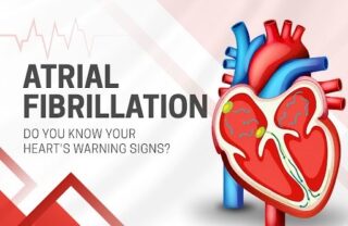
Blood Clots in AFib: How They Form and Prevention
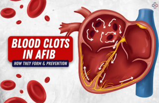
Blood Clots in AFib You don’t think about your blood clotting. You just assume it works the way nature intended.
Blood Clots in AFib You don’t think about your blood clotting. You just assume it works the way nature intended.
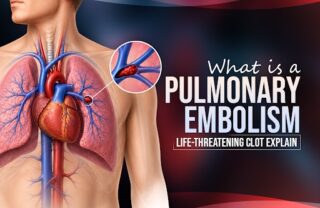
Pulmonary embolism You’re sitting at work when suddenly you can’t catch your breath. Your chest feels tight. Your heart is

CTEPH -You survived a blood clot in your lungs. That pulmonary embolism was terrifying—sudden shortness of breath, chest pain, panic.
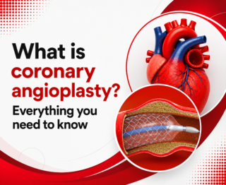
Coronary angioplasty: Your chest feels tight. You’ve been getting shortness of breath lately, especially when you climb stairs or walk
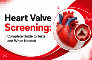
The Silent Problem You Might Not Know You Have Heart Valve Screening Heart Valve Screening. You’re sitting at your desk.

The Moment Your World Shifts Heart You’re sitting in your doctor’s clinic, discussing concerns with a heart specialist in Hyderabad.
You’re sitting at your desk, finishing that important presentation. Your heart suddenly feels like it’s doing flip-flops in your chest.

Cardiologist in Hyderabad are increasingly seeing cases like Rajesh’s. Rajesh, 40, a non-smoker and non-drinker, IT team lead in Hyderabad,

Heart doctor in Hyderabad are increasingly seeing a troubling trend among younger adults. You are 34, a non-smoker, not diabetic,
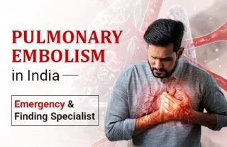
A Clot That Travels to the Lung and the Urgency It Demands Pulmonary embolism (PE) is a sudden blockage of
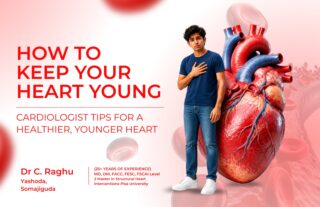
Cardiologist tips check your phone, skip breakfast, and rush through traffic to reach the office. After sitting for long hours,

Best heart doctor Comprehensive Cardiac Care — Why It Matters for the Patient Cardiac conditions do not present in isolation.
© 2024, Dr. Raghu. All rights reserved. Design & Developed by AMSDigital.in
+91 95424 75650
For Appointment with Dr Raghu:
+91 80085 36699
Dr Raghu’s Patient Coordinator:
+91 72860 10203
Dr Raghu’s Office Experience:
+91 90001 65962
Dr Raghu’s Social Media Communication Channel:
+91 97046 51708
