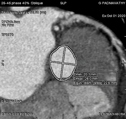
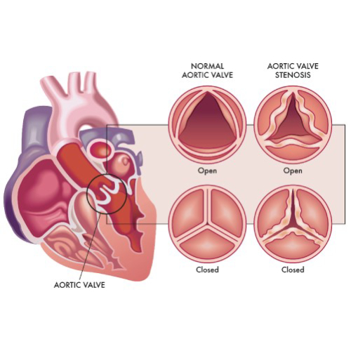
Transcatheter Aortic Valve Replacement (TAVR) has revolutionized the landscape of cardiovascular interventions, providing a less invasive alternative to traditional open-heart surgery for treating aortic valve stenosis.
aortic valve replacement | Dr Raghu
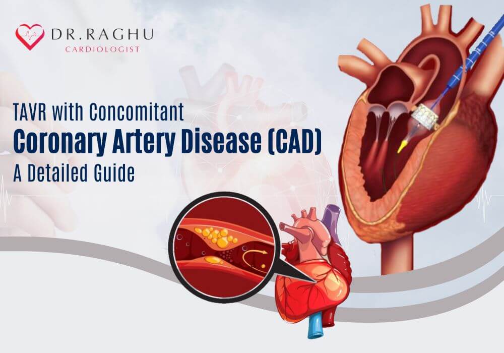
Transcatheter Aortic Valve Replacement (TAVR) has emerged as a revolutionary procedure in the field of cardiology, providing a less invasive alternative to traditional open-heart surgery for patients suffering from severe aortic valve stenosis. Recent advancements have expanded the applications of TAVR to include patients with concomitant coronary artery disease (CAD), presenting a groundbreaking approach to addressing multiple cardiac issues simultaneously.
In this article, we will take a closer look at the benefits and key considerations for TAVR with concomitant CAD.
Understanding TAVR
Transcatheter Aortic Valve Replacement (TAVR), also known as Transcatheter Aortic Valve Implantation (TAVI), is a medical procedure used to treat aortic valve stenosis. Aortic valve stenosis is a condition in which the aortic valve in the heart becomes narrowed, restricting the flow of blood from the heart to the rest of the body.
Traditionally, aortic valve replacement has been performed through open-heart surgery, involving the removal of the diseased valve and its replacement with a prosthetic valve. However, TAVR offers a less invasive alternative for certain patients, especially those who are considered high-risk or ineligible for open-heart surgery due to age or other health issues.
Concomitant Coronary Artery Disease and TAVR
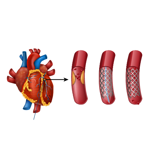
Concomitant Coronary Artery Disease (CAD) refers to the presence of atherosclerosis or plaque buildup in the coronary arteries alongside aortic stenosis. In the past, patients requiring both aortic valve replacement and coronary artery intervention would often undergo separate procedures. However, recent studies have explored the feasibility and efficacy of combining TAVR with percutaneous coronary intervention (PCI) in a single hybrid procedure.
Benefits of combining TAVR with concomitant CAD treatment include:
Reduced Procedural Burden
Combining TAVR with PCI in a single procedure reduces the overall procedural burden on the patient. This minimizes the need for multiple hospitalizations, lowers the risk of complications, and accelerates the overall recovery process.
Multifaceted Approach
TAVR with concomitant CAD allows physicians to address both aortic valve stenosis and coronary artery blockages in one session. This multifaceted approach enhances patient outcomes by comprehensively managing multiple cardiac issues simultaneously.
Minimized Anesthesia Exposure
Performing a single hybrid procedure limits the duration of anesthesia exposure for the patient, which can be particularly beneficial for elderly individuals or those with comorbidities who may be more sensitive to prolonged anesthesia.
Streamlined Postoperative Care
Streamlining interventions into a single procedure simplifies postoperative care, as patients only need to recover from one surgery. This may contribute to shorter hospital stays and a more efficient rehabilitation process.
Challenges and Considerations for TAVR With Concomitant CAD
While TAVR with concomitant CAD presents promising advantages, several challenges and considerations need to be addressed:
Patient Selection
Careful patient selection is crucial to ensure that individuals are suitable candidates for combined TAVR and PCI. Factors such as anatomical considerations, the complexity of coronary artery disease, and overall patient health must be thoroughly evaluated.
Technical Challenges
Performing both TAVR and PCI in a single procedure poses technical challenges that demand a high level of expertise from the interventional cardiology team. This includes precise coordination, imaging guidance, and a deep understanding of the complexities associated with both procedures.
Optimal Timing
Determining the optimal timing for TAVR with concomitant CAD remains a subject of ongoing research. Balancing the urgency of aortic valve replacement with the planning required for coronary intervention is crucial for achieving optimal outcomes.
Final Thoughts
The integration of TAVR with concomitant CAD marks a significant stride forward in the realm of cardiovascular interventions. This approach offers a promising solution for patients with multiple cardiac conditions, streamlining the treatment process and potentially improving overall outcomes. As ongoing research continues to refine patient selection criteria and procedural techniques, the future of TAVR with concomitant CAD holds great promise in revolutionizing the landscape of cardiovascular medicine.
Dr. C Raghu is regarded as the best cardiologist in Hyderabad. He specializes in interventional cardiology and has treated countless patients with severe heart ailments. If you or anyone you know is looking for TAVR surgery in Hyderabad, reach out to Dr. Raghu today.
Book Online Consultaion
TAVR With Concomitant Coronary Artery Disease (CAD): A Detailed Guide
Subscribe the Hearty Life Blogs

DR. RAGHU | Best Cardiologist in Hyderabad
Cardiology Coronary, Vascular and
Structural Interventions
Conditions & Diseases
Angioplasty
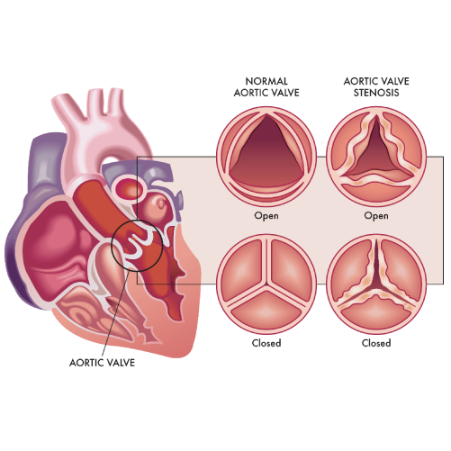
Aortic Stenosis
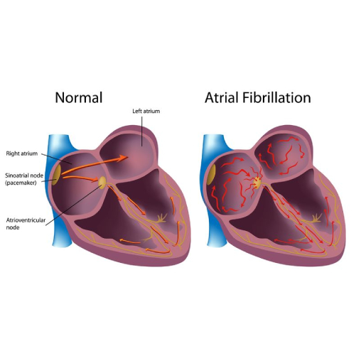
Atrial Fibrillation
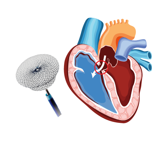
Atrial Septal Defect
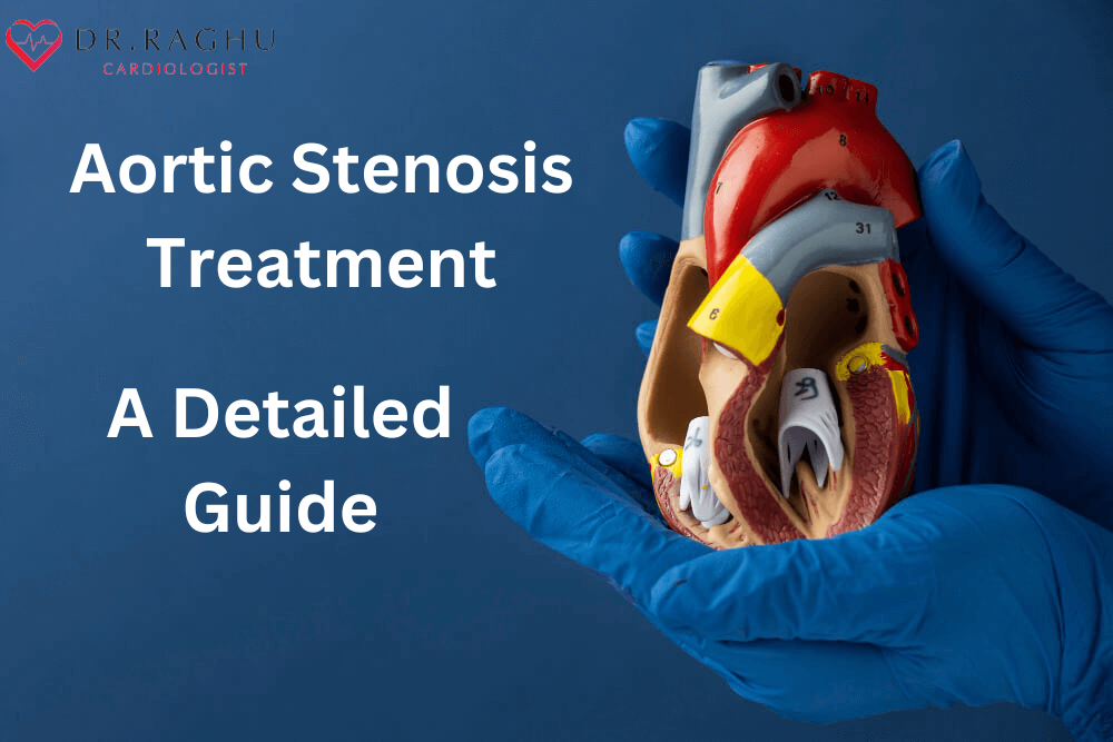
Aortic stenosis is a narrowing of the aortic valve, which restricts blood flow from the heart to the body. Treatment options include medication, balloon valvuloplasty, and aortic valve replacement. Learn more about aortic stenosis treatment and how to choose the right option for you.
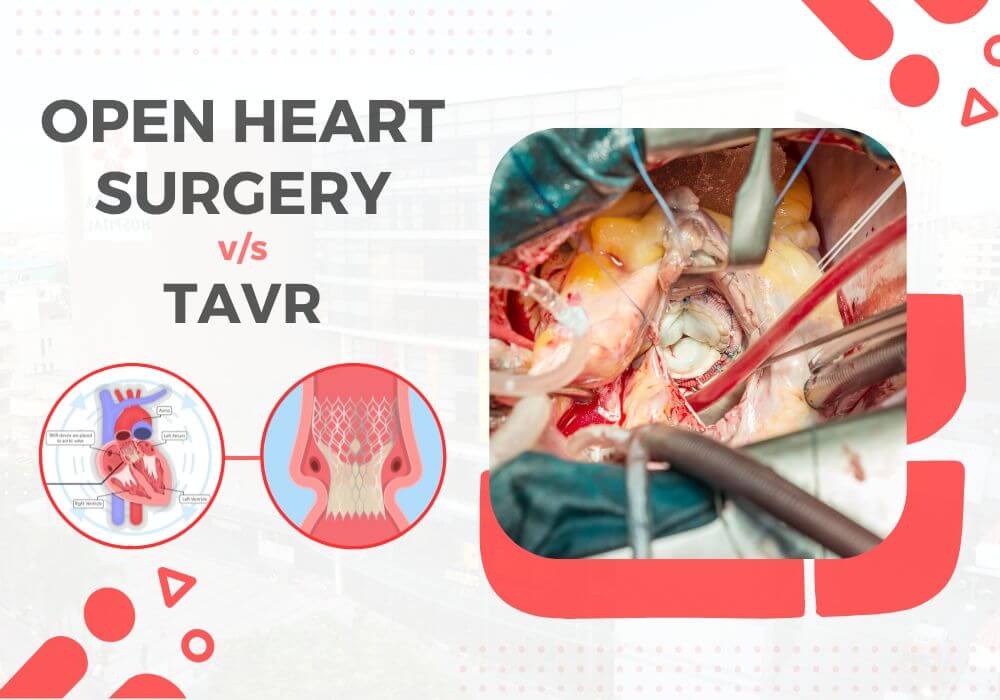
Open heart surgery and TAVR are two distinct approaches to treating aortic valve stenosis. Open heart surgery remains the gold standard for its durability and versatility in addressing complex cases. On the other hand, TAVR presents a less invasive alternative, particularly beneficial for high-risk patients and those who cannot undergo traditional surgery.
Before you undergo TAVR, your cardiologist will recommend various diagnostic tests to assess your overall health and risk factors. One of the most common tests that doctors prescribe is a CT angiogram.
Aortic valve stenosis is a serious condition that doesn’t just affect the aortic valve. It reduces or blocks the blood supply to the aorta. That, in turn, restricts blood flow to vital organs, including the brain, kidneys, and liver.
Grading Severity of Aortic Stenosis
Aortic valve stenosis is a serious disease that can lead to heart failure, strokes, and even death if left untreated. That makes timely diagnosis and treatment of the condition crucial. However, treatment of aortic stenosis depends on its severity.
Typically, the following parameters are used to determine the severity of aortic stenosis:
- Pressure gradient – High gradient (HG; >/=40 mm Hg) or low gradient (LG; <40 mm Hg)
- Blood flow – Normal flow (NF; SVi>35 ml/m2) or low flow (LF; SVi<35ml/m2)
- Left ventricular ejection fraction (LVEF) – Preserved (>/=50%) or reduced (<50%)
Depending on the pressure gradient and blood flow parameters, aortic stenosis is graded as follows:
- Normal flow-low gradient (NF-LG)
- Normal flow-high gradient (NF-HG)
- Low flow-high gradient (LF-HG)
- Low flow-low gradient (LF-LG)
NF-HG is the most prevalent type of aortic stenosis and has well-established management protocols. Patients with NF-HG are also ideal candidates for aortic valve replacement. While LF-LG is fairly rare, it’s often associated with a poor prognosis.
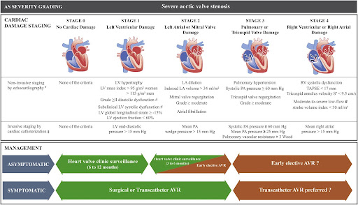
Additionally, depending on progression, heart valve disease can be categorized into the following four stages:
- Stage A (At risk) – Characterised by the presence of risk factors
- Stage B (Progressive) – Mild or moderate valve disease with no noticeable symptoms
- Stage C (Asymptomatic severe) – Severe valve damage with no noticeable symptoms
- Stage D (Symptomatic severe) – Severe valve disease with noticeable symptoms
Doctors use a variety of diagnostic tests to evaluate the aforementioned parameters and determine the severity of aortic stenosis. If you experience symptoms like chest pain, heart murmur, or palpitation, it’s crucial to reach out to an experienced cardiologist and get the right treatment for aortic valve stenosis.
How Does One Diagnose Aortic Stenosis – ECG, ECHO, TEE, or CT aortogram?
Early diagnosis of aortic valve stenosis is crucial to prevent severe complications, such as arrhythmias, heart failure, stroke, and death. Also, it can help administer timely treatment, thus improving the patient’s prognosis and quality of life.
That’s why cardiologists use a series of tests to diagnose aortic valve stenosis and its underlying cause. When you visit the doctor, they’ll start by asking you about your symptoms and medical history. Also, they ask whether your family has a history of cardiovascular ailments. Next, they’ll use a stethoscope to detect the presence of the characteristic aortic stenosis murmur.
Additionally, your doctor will use one or more of the following tests for the complete diagnosis:
- ECG (Electrocardiogram) – It’s one of the most preliminary tests that evaluate the heart’s electrical activity and helps doctors identify an irregular heartbeat and other abnormalities.
- Echo (Echocardiogram) – It uses sound waves to generate images of the beating heart. It helps doctors examine how blood flows through each valve and determine the severity of aortic stenosis.
- TEE (Transesophageal echocardiogram) – It’s a special type of echocardiogram in which an ultrasound probe is inserted into the esophagus and directed closer to the heart. It helps doctors take a closer look at the aortic valve and identify the underlying cause of aortic stenosis.
- CT aortogram – It’s used to evaluate the blood supply to the upper body and identify conditions such as atherosclerosis.
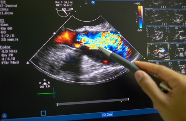
Additionally, your doctor might recommend tests like cardiac catheterization and chest X-ray to get a complete picture of your cardiac health and plan the right course of treatment.

