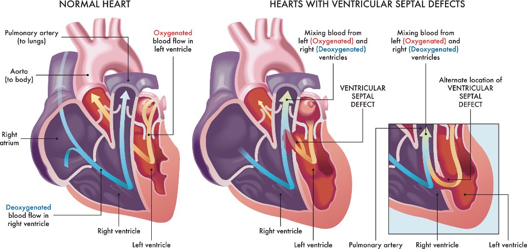
The heart consists of four chambers, of which upper two chambers are called as atria and lower two chambers are called as ventricles. Right and left chambers are separated by a wall of muscle called a septum. The septum separating the atria is called inter atrial septum and the once between the two ventricles in inter ventricular septum.
Right two chambers – right atrium and right ventricle pump the deoxygenated blood collected from various parts of the body to the lungs. Blood gets saturated with oxygen in the lungs and returns back to the left side of the heart.
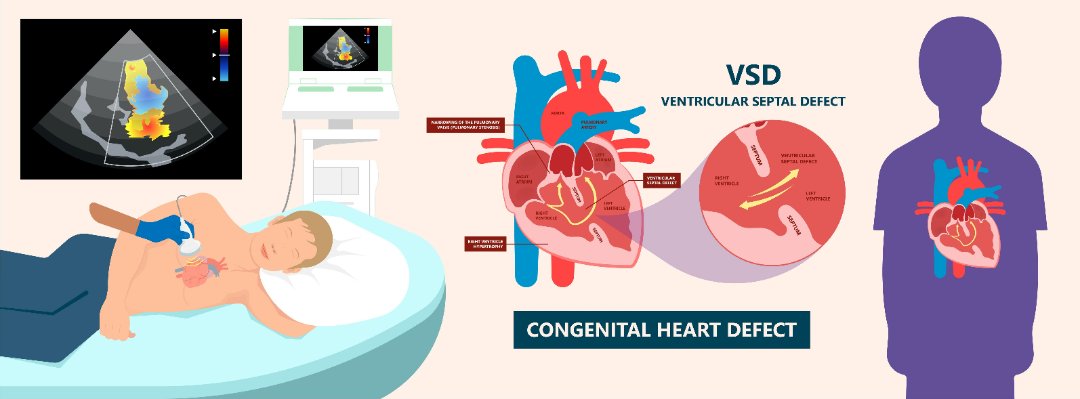
Ventricular septal defects (VSD) is a common type of congenital heart defect, which is characterized by an abnormal opening or a hole in the interventricular septum, the dividing wall between right and left ventricles. The oxygen-rich blood from the left ventricle will enter into the right ventricle through the opening, thereby getting mixed with deoxygenated blood which then enters into lungs. This will force the heart and lungs to work harder.
Causes:
The exact cause of VSD is unknown. During fetal heart developmental stage, the heart develops from a large tube which eventually divides into chambers and walls. Any abnormality in this process will lead to the formation of a defect in the septum. If the defect is in the interventricular septum, then it is said to be ventricular septal defect. There may be one or more VSDs.

Types:
Based on the location and development of VSD, it is classified into following types:
- Conoventricular Ventricular Septal Defect: It occurs just below the pulmonary and aortic valves.
- Perimembranous Ventricular Septal Defect: It occurs in the upper part of the ventricular septum.
- Inlet Ventricular Septal Defect: It occurs adjacent to the tricuspid and mitral valves. This type of defect might be associated with atrial septal defect.
- Muscular Ventricular Septal Defect: It is the most common type of VSD, which occurs in the lower muscular part of the interventricular septum.
Signs and symptoms:
Small defects in the septum do not show any symptoms because it closes on its own gradually during childhood.
Large defects show signs and symptoms usually after birth within a few weeks or months.
The first sign of VSD is heart murmur, which is a whooshing sound that can be heard using a stethoscope. The other symptoms include:
- Fatigue (tiredness)
- Arrhythmias (abnormal heart rhythm)
- Fast breathing or breathlessness
- Poor feeding
- Poor weight gain
- Pale skin
- Enlarged liver
Risk factors:
Ventricular septal defects mostly occur due to defective genes and chromosomes, that may be hereditary.
Environmental factors during pregnancy may also play a role in development of VSD in the fetus.
Complications:
Large or medium septal defects if left untreated, may lead to life threatening complications such as:
- Heart failure, as heart need to work harder to pump enough blood to the body.
- Pulmonary hypertension (increased blood flow to the lungs results in increased blood pressure in the lung arteries).
- Endocarditis (infection in the endocardium of heart).
- Other heart problems such as abnormal heart rhythms and valve problems.

Diagnosis:
If heart murmurs are detected during the physical examination, the patient may be advised further testing to conform the diagnosis.
- Echocardiogram: This test is the primary tool for the diagnosis of VSD, as it can be used to determine the size, location and severity of the ventricular septal defect. Sound waves are used to produce the detailed images of the heart.
- Electrocardiogram (ECG): This test records the electrical activity of the heart and helps to identify the abnormal heart rhythms and defects in the septum.
- Chest X-ray: X-rays are used to view the images of heart and lungs. In VSD, this test helps to determine the enlarged heart and extra fluid in the lungs.
- Cardiac catheterization: The test involves inserting a thin, flexible tube into the blood vessel at the groin or an arm, which is guided to the heart, to identify any congenital heart defects and to determine the function of the heart chambers and its valves.
- Pulse oximetry: Oxygen levels in the blood can be measured using a small clip which is placed on the fingertip.
Treatment:
Usually treatment is not needed for small VSDs, as they close on their own gradually after birth.
Babies with larger VSD need surgery to prevent any further complications.
The treatment for ventricular septal defects include:
- Medications such as diuretics like furosemide are used to reduce the amount of fluid in the blood that is pumped to the lungs and in the circulation. Beta blockers like metoprolol, propranolol and digoxin are used to maintain the regular heartbeat.
- Surgical procedures include surgical repair, catheter procedure and hybrid procedure.
Surgical repair involves open heart surgery, in which the doctor uses a patch or stitches to close the hole, performed under general anaesthesia.
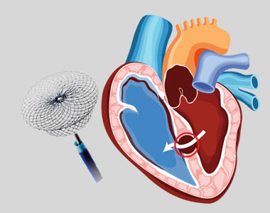
In catheter procedure, a catheter is inserted into a blood vessel and then passed to the heart. The defect is closed using a specially sized mesh device.
Hybrid procedure uses both surgical repair and catheterization procedures to close the hole, with the help of a heart-lung machine and a catheter placed through an incision.
Prevention:
Ventricular septal defects cannot be prevented, but following certain measures during pregnancy may be helpful to prevent the risk of VSD. The measures include:
- Prenatal care before pregnancy: Consult a physician before planning for pregnancy and inform him about the family history of any congenital defects and the medications using currently, so that he may give suggestions or recommend some lifestyle modifications to avoid the risk of heart defects.
- Healthy diet: Having a balanced diet including vitamins and folic acid during pregnancy will help in giving birth to a healthy child without any heart defects.
- Regular exercise under the supervision of a gynecologist is necessary during pregnancy.
- Avoid alcohol, tobacco and harmful drug use : during pregnancy to prevent the risk of VSD.
- Get vaccinated: Check whether you are vaccinated for vaccine-preventable infections before getting pregnant.
- Control diabetes: Monitor your sugar levels regularly to prevent the risk of heart defects.


