second-opinion-before-heart-valve-surgery.webp

August 26, 2026 AbhijitHearty Life

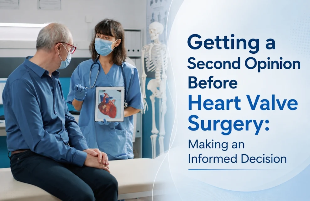
A heart valve diagnosis can create uncertainty for patients and their families. When heart valve surgery is recommended, patients may have questions about the diagnosis, available procedures, surgical risks, recovery, and whether another treatment option may be appropriate. Seeking a second medical opinion can provide additional clarity and help patients make an informed decision based on their individual condition.

A second opinion does not necessarily mean that the first recommendation is incorrect. It offers an opportunity to review the diagnosis, understand the severity of the valve problem, and discuss whether surgical or catheter-based treatment is suitable.

Why Consider a Second Opinion Before Heart Valve Surgery?

 heart valve surgery

Heart valve disorders can vary considerably from one patient to another. The appropriate treatment depends on factors such as the affected valve, severity of disease, symptoms, age, heart function, other medical conditions, and previous cardiac procedures.

Before proceeding with heart valve surgery, a second opinion may help clarify:

  • Whether the diagnosis is accurate and complete
  • How severe the valve disease is
  • Whether treatment is required immediately or can be monitored
  • Whether open heart surgery is necessary
  • Whether a minimally invasive or catheter-based procedure may be suitable
  • What recovery may involve
  • What the expected benefits and potential risks of treatment are

This additional evaluation can be particularly relevant for patients who have been advised to undergo complex valve procedures.

Understanding Different Heart Valve Treatment Options

The term “heart valve treatment” includes several approaches. Traditional surgical valve repair or replacement may be appropriate for certain patients, while others may qualify for catheter-based interventions.

For patients with severe aortic stenosis, Transcatheter Aortic Valve Replacement (TAVR) may be considered in appropriate circumstances. TAVR involves replacing the diseased aortic valve through a catheter rather than conventional open-heart surgery.

Patients should not assume that TAVR or surgery is automatically better. A specialist assessment is necessary to determine the most appropriate treatment based on clinical findings and imaging results.

When Is TAVR Considered?

TAVR is primarily used for selected patients with significant aortic valve disease, particularly aortic stenosis. Factors such as age, surgical risk, anatomy, valve characteristics, associated diseases, and overall health can influence treatment selection.

A consultation with a TAVR expert in Hyderabad can help eligible patients understand whether transcatheter valve replacement may be an option. A TAVR expert may evaluate echocardiography, CT imaging, cardiac catheterization findings, and other clinical information before recommending a treatment pathway.

The decision should be individualized rather than based solely on age or a single test result.

Second Opinion for Mitral Valve Disease

second opinion mitral valve disease

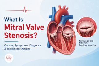
Mitral valve disease may also require careful evaluation. Mitral stenosis, for example, can restrict blood flow through the heart and may produce symptoms such as breathlessness, fatigue, or reduced exercise tolerance.

Depending on the severity and anatomy of the valve, treatment options can include medication for symptom management, balloon procedures, valve repair, or surgery.

Patients seeking mitral valve stenosis treatment may benefit from discussing all appropriate options with a qualified specialist. In selected cases, balloon mitral valvotomy may be considered, while other patients may require mitral valve surgery.

The choice depends on detailed clinical and imaging assessment.

What Should Patients Take to a Second-Opinion Consultation?

A second-opinion consultation becomes more useful when the specialist has access to the patient’s complete medical information. Patients should ideally carry:

  • Echocardiography reports and images
  • CT or MRI reports, where applicable
  • Coronary angiography or catheterization records
  • Previous medical and surgical history
  • Current medication list
  • Blood test reports
  • Previous hospital discharge summaries
  • Details of symptoms and their progression

These records allow the specialist to review the case more comprehensively rather than relying only on a written diagnosis.

Choosing the Right Specialist

Patients should consider consulting a cardiologist or cardiac specialist with experience relevant to their specific valve condition. A cardiologist in Hyderabad can assess the cardiovascular aspects of the condition, while patients requiring procedural or surgical evaluation may need consultation with an appropriately trained cardiac intervention or surgical team.

For patients researching a heart surgeon in Hyderabad, it is important to consider the specialist’s experience with the specific valve disorder and the treatment options being considered. the best cardiologist

Patients may also search for the best cardiologist in Hyderabad or Best cardiologist in Somajiguda, but treatment decisions should be based on clinical expertise, appropriate evaluation, and the individual patient’s medical needs rather than rankings or promotional claims.

Questions to Ask During a Second Opinion

Patients can make the consultation more productive by asking direct questions, such as:

  1. What type and severity of valve disease is present?
  2. Is heart valve surgery currently necessary?
  3. Can the valve be repaired, or does it require replacement?
  4. Is catheter-based treatment an option?
  5. Would TAVR/TAVI be appropriate in this case?
  6. What are the potential benefits and risks of each treatment?
  7. What could happen if treatment is delayed?
  8. What type of follow-up and recovery will be required?

Understanding these points can help patients and families participate more confidently in treatment decisions.

Also read: Why Get a Second Opinion on Heart Problems? When & How 

Making an Informed Heart Valve Treatment Decision

A second opinion is an important part of informed decision-making when major cardiac treatment is being considered. It can help patients understand their diagnosis, compare suitable treatment approaches, and clarify why a particular procedure has been recommended.

For conditions ranging from aortic stenosis to mitral valve disease, heart valve disease treatment should be individualized according to clinical findings, imaging, symptoms, overall health, and procedural suitability.

Patients considering heart valve surgery, TAVR, or other valve interventions can seek a detailed evaluation from Dr. C. Raghu, Clinical Director & Senior Interventional Cardiologist in Hyderabad, for an informed discussion about appropriate treatment options.

If you or a family member has been advised to undergo heart valve treatment, consult Dr. C. Raghu for a comprehensive cardiac evaluation and second opinion before making an important treatment decision.


WhatsApp-Image-2026-08-17-at-15.29.37.jpeg

August 17, 2026 AbhijitHearty Life

Biomarkers in ACS play an important role in identifying heart muscle injury and supporting the diagnosis and risk assessment of acute coronary syndrome (ACS). Among the available cardiac biomarkers, cardiac troponin—particularly high-sensitivity cardiac troponin (hs-cTn)—is central to the modern evaluation of patients with suspected ACS.

What Is Acute Coronary Syndrome?

Acute coronary syndrome is a group of conditions caused by a sudden reduction in blood flow to the heart muscle. It includes unstable angina, non-ST-segment elevation myocardial infarction (NSTEMI), and ST-segment elevation myocardial infarction (STEMI).

Symptoms may include chest discomfort, breathlessness, sweating, nausea, or pain spreading to the arm, shoulder, jaw, or back. Because symptoms can vary, clinical assessment is combined with an electrocardiogram (ECG) and blood tests.

What Are Biomarkers in ACS?
High-Sensitivity Troponin Important

Biomarkers in ACS are measurable substances in the blood that can provide evidence of myocardial injury. The most clinically important biomarker is cardiac troponin, a protein released into the bloodstream when heart muscle cells are damaged.

The two major forms used clinically are cardiac troponin I and cardiac troponin T. Modern high-sensitivity cardiac troponin assays can detect much smaller concentrations of troponin than earlier tests, allowing clinicians to identify myocardial injury earlier and more accurately.

However, an elevated troponin concentration does not automatically mean that a patient has had a heart attack. Troponin can also rise in conditions such as heart failure, myocarditis, pulmonary embolism, kidney disease, severe infection, or sustained rapid heart rhythms. Therefore, results must be interpreted alongside symptoms, ECG findings, medical history, imaging, and changes in troponin levels over time.

Why Is High-Sensitivity Troponin Important?

High-sensitivity cardiac troponin has become an important component of acute coronary syndrome diagnosis. It can detect low concentrations of cardiac troponin and help clinicians identify or rule out myocardial infarction more efficiently.

Instead of relying only on a single blood test, clinicians commonly assess the troponin concentration at presentation and, when appropriate, repeat it after a defined interval. The change between measurements—often referred to as the delta troponin—can provide additional information about an acute process.

This approach is particularly relevant to the diagnosis of NSTEMI, where there may be no persistent ST-segment elevation on the ECG.

High-Sensitivity Troponin Algorithms

High-sensitivity troponin algorithms use specific hs-cTn measurements and changes over time to help classify patients with suspected ACS. Depending on the assay and clinical setting, protocols may use very early measurements followed by repeat testing after a short interval.

A troponin algorithm can help place patients into broad clinical categories, such as:

  • Patients in whom acute myocardial infarction can be ruled out.
  • Patients who require additional observation and testing.
  • Patients with findings that support an acute myocardial infarction and require urgent management.

The exact thresholds and timing vary according to the specific assay, laboratory, clinical protocol, and patient characteristics. Therefore, hs-cTn results should not be interpreted independently.

Troponin for Acute Coronary Syndrome

Troponin for acute coronary syndrome is valuable because it provides objective evidence of myocardial injury. A rise and/or fall in cardiac troponin, together with clinical evidence of myocardial ischemia, is central to diagnosing acute myocardial infarction.

It is important to distinguish myocardial injury from myocardial infarction. An elevated hs-cTn level indicates myocardial injury, while myocardial infarction requires evidence that the injury is related to myocardial ischemia.

This distinction helps prevent overdiagnosis and ensures that patients receive an appropriate evaluation for the underlying cause.

Other ACS Biomarkers

Although cardiac troponin is the preferred biomarker for suspected myocardial infarction, researchers have studied several other ACS biomarkers, including creatine kinase-MB and myoglobin.

Creatine kinase-MB has historically been used to detect myocardial injury but is less specific than cardiac troponin. Myoglobin can rise early after muscle injury but lacks cardiac specificity. For these reasons, contemporary clinical practice generally places cardiac troponin at the center of biomarker-based assessment.

How Are Biomarkers Used With Other Tests?

A reliable acute coronary syndrome diagnosis does not depend on biomarkers alone. Healthcare professionals consider several factors together:

  1. Symptoms and medical history – The nature, duration, and associated features of chest discomfort are assessed.
  2. ECG – An ECG can identify ischemic changes and may immediately indicate STEMI.
  3. hs-cTn testing Initial and repeat measurements help assess myocardial injury.
  4. Clinical examination: Blood pressure, heart rate, oxygenation, and other findings provide additional information.
  5. Imaging and coronary evaluation: Echocardiography, coronary CT angiography, or invasive coronary angiography may be considered when clinically appropriate.

This combined approach is particularly important because a normal initial troponin result does not always exclude ACS when testing occurs very early after symptom onset.

When Should Medical Attention Be Sought?

New or unexplained chest pressure, severe breathlessness, fainting, cold sweating, or discomfort spreading to the arm, jaw, shoulder, or back may indicate a serious cardiac problem. Such symptoms require urgent medical assessment rather than relying on an online explanation or waiting for symptoms to resolve.

Early evaluation can allow clinicians to perform an ECG and appropriate blood tests, including hs-cTn, and determine whether further emergency treatment is required.

Conclusion

Biomarkers in ACS, particularly hs-cTn, have transformed the assessment of suspected acute coronary syndrome. High-sensitivity cardiac troponin allows clinicians to detect myocardial injury at lower concentrations and use structured high-sensitivity troponin algorithms to support rapid clinical decision-making.

Nevertheless, troponin results must always be interpreted in context. Symptoms, ECG findings, serial measurements, medical history, and other investigations remain essential for accurate diagnosis and appropriate treatment.

For expert evaluation of suspected coronary disease or acute cardiac symptoms, consultation with a qualified specialist such as the best cardiologist in Hyderabad can help ensure timely and appropriate cardiac assessment.

For cardiac consultation and evaluation, contact Dr. C. Raghu, Clinical Director & Senior Interventional Cardiologist at Yashoda Hospitals, Secunderabad.

Click here for more information on  YouTube: https://youtu.be/zhvamAmo3JA?si=sV8GaWWPjUA8Cjhj

Important: This article on biomarkers in ACS is intended for general educational and informational purposes only and does not replace professional medical advice, diagnosis, or treatment. If you or someone around you develops symptoms suggestive of a heart attack, such as chest pain or pressure, severe breathlessness, sweating, nausea, or pain spreading to the arm, jaw, shoulder, or back, seek emergency medical care immediately.


August 12, 2026 AbhijitHearty Life

If breathlessness on stairs, fatigue that won’t lift, or a racing heartbeat has sent you searching for answers, and a doctor has mentioned  balloon mitral valvotomy and mitral valve stenosis in the same sentence, you’re likely trying to understand balloon mitral valvotomy for your daily life  not just your test report. This article breaks down what balloon mitral valvotomy is, who it helps, what the procedure and recovery look like, and the questions worth asking your cardiologist before deciding.

What Is Mitral Valve Stenosis?
Mitral valve stenosis

The mitral valve sits between the heart’s left atrium and left ventricle, controlling blood flow between the two chambers. In mitral valve stenosis, this valve narrows and stiffens, most often due to rheumatic heart disease from an earlier, sometimes unnoticed, episode of rheumatic fever. As the opening narrows, the heart has to work harder to push blood through, which can lead to breathlessness, fatigue, palpitations, and, in advanced cases, fluid buildup in the lungs or legs.

Because symptoms often develop gradually, many patients live with a narrowing valve for years before it’s picked up frequently during a routine check-up or an echocardiogram done for an unrelated reason.

What Is Balloon Mitral Valvotomy?

balloon mitral valvotomy

Balloon mitral valvotomy—also called percutaneous balloon mitral valvotomy or PBMV—is a catheter-based procedure used to widen a narrowed mitral valve without open-heart surgery. A thin catheter with a deflated balloon at its tip is guided through a vein, typically from the groin, up into the heart and across the mitral valve. Once positioned, the balloon is inflated to gently separate the fused valve leaflets, improving blood flow.

It’s performed under local anesthesia with the patient conscious, usually takes a couple of hours, and is done in a cardiac catheterization lab rather than an operating theater.

Also read: What is CTEPH? Rare Disease Explained Simply.

Why It’s Often Preferred Over Open-Heart Surgery

For eligible patients, percutaneous balloon mitral valvotomy offers a meaningfully different recovery experience compared to surgical valve repair or replacement:

  • No large chest incision or use of a heart-lung machine
  • Shorter hospital stay in most cases
  • Faster return to routine activities
  • Lower procedural risk profile for suitable candidates
  • The valve is preserved rather than replaced

This doesn’t mean balloon mitral valvotomy suits everyone — valve anatomy, calcium deposits, and the degree of regurgitation all factor into eligibility, which is exactly why a detailed pre-procedure evaluation matters.

Who Is a Good Candidate for Balloon Mitral Valvotomy?

Not every case of mitral valve stenosis calls for balloon mitral valvotomy. Cardiologists typically assess candidacy using echocardiographic scoring systems that look at leaflet mobility, thickness, calcification, and involvement of the valve’s supporting structures. Patients with pliable, minimally calcified valves and no significant mitral regurgitation tend to be the best candidates.

Balloon mitral valvotomy is commonly considered for:

  • Patients with moderate to severe mitral valve stenosis causing symptoms
  • Younger patients who want to avoid or delay valve replacement
  • Pregnant women with symptomatic stenosis, where surgery carries added risk
  • Patients who are poor candidates for open-heart surgery due to other health conditions

If the valve is heavily calcified, badly scarred, or accompanied by significant regurgitation, valve repair or replacement surgery may be the more durable option  something your cardiologist will weigh against percutaneous balloon mitral valvotomy during evaluation.

Also Read: What is chronic total occlusion? Under Chip CTO angioplasty

What to Expect: Procedure and Recovery

On the day of the procedure, patients are usually awake but sedated, with local anesthesia at the catheter insertion site. Real-time imaging, often a combination of fluoroscopy and echocardiography, helps guide the catheter and balloon precisely across the valve.

After the procedure, most patients are monitored for a short period and can typically go home within a day or two, depending on their overall condition and how the heart responds. Recovery is generally faster than after open-heart surgery, with many people resuming normal activity within a week, though your cardiologist will set the exact timeline based on your case.

Follow-up echocardiograms are important afterward to confirm the valve is functioning well and to monitor for restenosis, which can occur over time in some patients.

Living With Mitral Valve Stenosis: Beyond the Procedure

Whether or not balloon mitral valvotomy is the right path, ongoing care matters. Patients with a history of rheumatic fever may need long-term antibiotic prophylaxis to prevent recurrence. Regular monitoring, awareness of symptom changes, and coordination with a cardiologist familiar with structural heart disease all play a role in long-term outcomes.

This is also where continuity of care counts — working with an experienced cardiologist in Hyderabad who understands structural valve disease, alongside coronary artery disease specialists and congenital aortic stenosis specialists in Hyderabad, can make follow-up decisions clearer, especially if symptoms evolve or a second opinion is needed down the line. Patients across the country often look for a trusted best cardiologist in India who can offer this kind of integrated, long-term valve care rather than a one-time consultation.

Questions Worth Asking Your Cardiologist

Before deciding on balloon mitral valvotomy versus surgery, it helps to walk into that conversation prepared:

  • Is my valve anatomy suitable for balloon valvotomy, based on imaging?
  • What are the specific risks in my case?
  • How long is a typical recovery for someone in my situation?
  • What are the chances the valve narrows again later, and how would that be managed?
  • What symptoms should prompt me to call sooner rather than wait for my next appointment?

The Bottom Line

Balloon mitral valvotomy has given many patients with mitral valve stenosis a way to feel better and stay active without the recovery burden of open-heart surgery  but it’s not a one-size-fits-all answer. The right choice depends on your specific valve anatomy, symptoms, and overall health, which is why a thorough evaluation with an experienced cardiology team is the real starting point, not the procedure itself. Many patients researching the top 5 cardiologists in Hyderabad are simply trying to find someone they can trust with exactly this kind of decision.

If you’ve been told you have mitral valve stenosis, don’t wait on the breathlessness or fatigue to get worse  book a consultation with Dr. C. Raghu to get a detailed valve assessment and find out whether balloon mitral valveotomy is the right fit for you.


Occlusion-Myocardial-Infarction-Symptoms-Treatment-1-1200x778.jpeg

August 3, 2026 AbhijitHearty Life

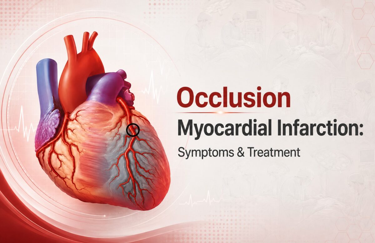
Occlusion myocardial infarction is a serious heart condition that occurs when a coronary artery becomes completely or significantly blocked, reducing or stopping blood flow to part of the heart muscle. Because prolonged loss of blood supply can cause permanent heart muscle damage, recognizing warning signs and getting emergency medical care quickly is extremely important.

For patients in Hyderabad, understanding the symptoms and treatment options can help them respond appropriately when a heart attack is suspected.

What Is Occlusion Myocardial Infarction?

A myocardial infarction, commonly called a heart attack, occurs when blood flow to the heart muscle is interrupted. In an occlusion myocardial infarction, a coronary artery may become completely blocked, often because a blood clot forms over a ruptured or damaged cholesterol plaque.

When oxygen-rich blood cannot reach the heart muscle, the affected tissue begins to become injured. The longer the artery remains blocked, the greater the potential for permanent heart damage.

This is why suspected heart attack symptoms should never be ignored or treated as simple indigestion, acidity, fatigue, or anxiety.

Common Symptoms of Occlusion Myocardial Infarction

Symptoms can vary between individuals, but common warning signs include:

  • Pressure, tightness, heaviness, or pain in the chest
  • Pain spreading to the arm, shoulder, back, neck, or jaw
  • Shortness of breath
  • Cold sweating
  • Nausea or vomiting
  • Unusual weakness or extreme tiredness
  • Dizziness or light-headedness
  • Rapid or irregular heartbeat
  • A feeling of anxiety or impending danger

Some people, particularly older adults and those with diabetes, may experience less typical symptoms. Chest discomfort may also be mild rather than severe.

If symptoms suggest a heart attack, seeking emergency medical attention immediately is more important than trying to identify the exact cause at home.

What Causes a Coronary Artery Occlusion?

Several factors can contribute to coronary artery blockage. One of the most common causes is coronary artery disease, where fatty deposits gradually accumulate inside the arteries.

Risk factors include:

  • High cholesterol
  • High blood pressure
  • Diabetes
  • Smoking or tobacco use
  • Obesity
  • Physical inactivity
  • Unhealthy diet
  • Family history of heart disease
  • Increasing age
  • Chronic stress
  • Previous cardiovascular disease

A plaque inside a coronary artery can sometimes rupture suddenly. The body may respond by forming a blood clot, which can rapidly restrict or completely stop blood flow.

How Is Occlusion Myocardial Infarction Diagnosed?

Doctors use several tests to determine whether a patient is experiencing a heart attack and to understand the severity of the blockage.

Electrocardiogram (ECG)

An ECG records the heart’s electrical activity and can identify patterns associated with acute coronary artery blockage.

Blood Tests

Cardiac biomarkers, particularly troponin, can help determine whether heart muscle injury has occurred.

Echocardiogram

An echocardiogram uses ultrasound to evaluate heart structure and function. It can help doctors identify areas of the heart that are not contracting normally.

Coronary Angiography

Coronary angiography allows doctors to examine the coronary arteries and identify significant narrowing or blockage. It can also help guide treatment decisions.

Treatment for Occlusion Myocardial Infarction

Treatment depends on the patient’s condition, the location and severity of the blockage, and how quickly medical care is provided.

Emergency Medical Treatment

Doctors may use medications to reduce clot formation, improve blood flow, control pain, and stabilize the patient’s condition.

Coronary Angioplasty and Stenting

For an acutely blocked coronary artery, percutaneous coronary intervention (PCI) may be performed. During the procedure, a catheter is guided into the affected artery. A balloon can be used to open the narrowed area, and a stent may be placed to help maintain blood flow.

Rapid restoration of blood flow is an important goal in appropriate patients with an acute coronary artery occlusion.

Additional Treatment

Depending on the patient’s cardiovascular risk and overall health, treatment may also include medicines for cholesterol, blood pressure, blood clot prevention, and other underlying conditions.

After the acute event, lifestyle changes and cardiac rehabilitation may play an important role in reducing future cardiovascular risk.

Also read:  What is chronic total occlusion? Under Chip CTO angioplasty  

Why Time Matters During a Heart Attack

Heart muscle requires a continuous supply of oxygen-rich blood. When a coronary artery becomes blocked, the affected muscle can progressively become damaged.

For this reason, people experiencing possible heart attack symptoms should not wait for symptoms to disappear on their own. Delaying medical evaluation can reduce the opportunity for timely treatment.

If someone develops severe or persistent chest discomfort, breathlessness, sweating, fainting, or pain spreading to the arm, jaw, back, or shoulder, emergency medical care should be sought immediately.

Choosing a Heart Specialist in Hyderabad

Patients dealing with suspected or diagnosed coronary artery disease may need evaluation by an experienced cardiologist in Hyderabad. A heart specialist in Hyderabad can assess cardiovascular risk factors, interpret diagnostic tests, and recommend an appropriate treatment strategy.

For patients with complex coronary conditions, specialist evaluation can help determine whether medication, coronary angioplasty, stenting, or another intervention is appropriate.

Searching for a myocardial cardiologist in Hyderabad may also lead patients to specialists who manage heart attacks and other coronary artery conditions.

Can Occlusion Myocardial Infarction Be Prevented?

Not every heart attack can be prevented, but many cardiovascular risk factors can be managed.

Helpful measures include:

  • Controlling blood pressure and cholesterol
  • Managing diabetes effectively
  • Avoiding smoking and tobacco
  • Maintaining a healthy body weight
  • Following a heart-healthy diet
  • Exercising according to medical advice
  • Taking prescribed medicines regularly
  • Attending recommended cardiovascular check-ups
  • Recognizing and acting on warning symptoms promptly

People with known heart disease or multiple cardiovascular risk factors should discuss an appropriate prevention plan with their doctor.

Read more: What Is Atrial Fibrillation? Understanding AFib Simply 

When Should You Seek Emergency Care?

Chest pain or discomfort should never be ignored when it is sudden, severe, persistent, or accompanied by symptoms such as breathlessness, sweating, nausea, dizziness, or pain radiating to the arm or jaw.

Do not wait for symptoms to become severe before seeking emergency medical attention.

Occlusion myocardial infarction can become life-threatening when blood flow to the heart is significantly interrupted. Early recognition, rapid diagnosis, and timely treatment can make a critical difference.

Consult Dr. C. Raghu

If you are concerned about coronary artery disease, heart attack symptoms, or other cardiovascular problems, consultation with an experienced cardiology specialist can help with appropriate evaluation and treatment planning.

Dr. C. Raghu is a senior interventional cardiologist associated with advanced cardiac care in Hyderabad and Secunderabad.

Watch the video to review the discussion on occlusive myocardial infarction and acute coronary syndrome: https://youtu.be/jWvP0BG2bsY

For more information about cardiac care and interventional cardiology services,

Important: This article is intended for general educational purposes and does not replace professional medical advice. If you or someone around you has symptoms suggestive of a heart attack, seek emergency medical care immediately.


what-is-atrial-fibrillation.jpg

July 27, 2026 AbhijitHearty Life

Atrial fibrillation, commonly called AFib, is becoming increasingly common among middle-aged individuals. But here’s the thing—most people don’t understand what’s actually happening inside their hearts when this occurs. You might hear the term “AFib” from your doctor, but you’re left wondering what it really means for your life, your health, and your future.

What Exactly Happens During Atrial Fibrillation?

atrial fibrillation treatment

Let me explain this simply. Your heart works like a coordinated team. It has four chambers, and they’re supposed to contract in a specific rhythm, pumping blood efficiently throughout your body. When everything works correctly, your heart beats between 60 and 100 times per minute, and each beat is regular like a drum in an orchestra.

But with atrial fibrillation, something goes wrong. The two upper chambers of your heart, called the atria, start beating chaotically. Instead of contracting in a coordinated way, they’re essentially quivering. This irregular electrical activity causes your heart to beat irregularly and often rapidly. You might feel your heart skipping beats or racing without reason.

The problem is this: when your heart isn’t pumping efficiently, it doesn’t move blood through your body the way it should. This is serious.

Also read: What is coronary angioplasty? Everything you need to know  

Why Should You Care About This?

You might think, “Okay, so my heart beats irregularly. What’s the worst that can happen?” Here’s where you need to pay attention:

Blood clots form. When blood sits in your heart chambers instead of being pumped out efficiently, it can pool and clot. These clots can travel to your brain and cause a stroke—a brain stroke that could leave you paralyzed, unable to speak, or worse.

Your heart weakens. Over time, the constant irregular beating strains your heart muscle. It’s like running a machine non-stop without proper maintenance. Eventually, it breaks down, leading to heart failure.

You feel terrible. Shortness of breath, fatigue, dizziness—these aren’t just inconveniences. They affect your quality of life, your work, and your ability to enjoy time with your family.

Other complications arise. Your risk of heart disease increases. Your blood pressure becomes difficult to control. Your overall health deteriorates.

Who Gets Atrial Fibrillation?

You might be at higher risk if you:

  • Have high blood pressure
  • Are over 65 years old (though it can happen when younger)
  • Have heart disease or a previous heart attack
  • Have thyroid problems
  • Are obese or overweight
  • Drink excessive alcohol
  • Have sleep apnea
  • Experience chronic stress
  • Have family history of AFib

If any of these apply to you, you should be paying attention to any unusual heart sensations.

Atrial Fibrillation Treatment: What Are Your Options?

The good news? Atrial fibrillation is treatable. You’re not helpless here. You have options, and finding the right approach matters tremendously.

First, there’s medication. Your doctor might prescribe medications that help regulate your heart rate or convert your heart back to a normal rhythm. Some people do very well with medication alone.

Second, there are lifestyle changes. You need to reduce stress, exercise regularly (as approved by your doctor), limit alcohol, maintain a healthy weight, and manage any underlying conditions like high blood pressure.

Third, there are medical procedures. If medication doesn’t work, procedures like ablation can be performed. An interventional cardiologist specialist trained in heart procedures can access your heart and remove the tissue causing the irregular signals. It’s minimally invasive, meaning small incisions, less pain, and faster recovery.

Finding the Right Atrial Fibrillation Doctor in Hyderabad

If you’re in Hyderabad, you’re fortunate. The city has some of India’s finest cardiac specialists. When looking for an atrial fibrillation doctor in Hyderabad, you want someone experienced, compassionate, and thorough. You want a doctor who explains things clearly, who listens to your concerns, and who works with you to create a treatment plan that fits your life.

The best atrial fibrillation doctors in India understand that AFib isn’t just a medical condition—it’s something that affects how you live. They know that you want to return to your normal activities, spend time with your loved ones, and not feel like your health is controlling your life.

Taking Action Today

Don’t wait until you have a major event to address AFib. If you experience irregular heartbeats, shortness of breath, or persistent fatigue, schedule an appointment with a cardiologist. Get evaluated. Get answers.

Your heart is the most important muscle in your body. It’s literally keeping you alive. Treating atrial fibrillation early, with the guidance of a skilled cardiologist for atrial fibrillation, can prevent serious complications like brain stroke and significantly improve your quality of life.

Also read: TAVR/TAVI Procedure in Hyderabad Minimally Invasive Care  

Moving Forward

You don’t have to live with the fear and uncertainty of AFib. Modern medicine has given us excellent tools to manage this condition. The first step is getting a proper diagnosis and treatment from qualified specialists. The second step is taking responsibility for your health—making lifestyle changes, taking medications as prescribed, and maintaining regular follow-ups with your doctor.

Ready to take control of your heart health? Dr. C. Raghu, an experienced interventional cardiologist at Yashoda Hospital, Somajiguda, Hyderabad. Your heart deserves expert care.


chronic-total-occlusion.webp

July 23, 2026 AbhijitHearty Life

Chip cto angioplasty You’ve had blockages in your arteries for a while. You’ve had procedures. You’ve taken medications. And yet your doctor just told you that you have a completely blocked artery—what’s called a chronic total occlusion. The artery isn’t just narrowed. It’s not just severely blocked. It’s completely shut off.

Your first thought is panic. But before you despair, understand this: CTO doesn’t mean you’re out of options. It doesn’t mean your case is hopeless. Modern medicine has developed specialized techniques to open even completely blocked coronaries. But you need to understand what CTO is and how it’s treated.

Understanding Coronary Artery Disease Progression

Remember how coronary artery disease develops? Plaque builds up in your arteries over time. This usually happens over years or decades.

In some people, this process is gradual. The blockage slowly increases from 50% to 75% to 90%. At each stage, blood can still squeeze through, though less efficiently.

But sometimes, the blockage continues increasing until the artery is completely sealed. No blood flows through at all. This is a chronic total occlusion—or CTO.

The word “chronic” is important. It means this blockage didn’t just happen. It’s been there for a while—usually at least three months. Your body has actually adapted somewhat, sometimes developing other small blood vessels that partially compensate for the blocked artery.

Why CTO Causes Problems

Chronic Total Occlusion Treatment

You might think, “If blood develops alternative routes, why is CTO a problem?”

Here’s why it matters:

The alternative blood vessels are limited. They don’t fully replace the original artery’s function.

Your heart remains oxygen-starved. You still experience angina (chest pain) during exertion. You still feel fatigued.

Your heart remains vulnerable. If another artery narrows, your heart has fewer reserves.

It contributes to heart failure. Prolonged oxygen deprivation weakens your heart muscle.

It increases heart attack risk. The blocked artery region is particularly vulnerable to sudden deterioration.

Also read:  Blood Clots in AFib: How They Form and Prevention 

The Challenge of Chip CTO Angioplasty Treatment

Why don’t doctors simply reopen CTOs in everyone? Because it’s technically difficult.

When an artery is completely blocked, your doctor can’t thread a wire through the blockage the way they do with partially blocked arteries. The wire hits a dead end and can’t advance.

Recanalization—opening a completely blocked artery—requires specialized expertise, specialized equipment, and particular skill. Not all interventional cardiologists perform CTO procedures. It’s a specialized subset within the specialty.

CTO Angioplasty: How It Works

If you’re a good candidate for CTO angioplasty, here’s what happens:

Step One: Imaging Your doctor performs coronary angiography to visualize the completely blocked artery. They determine the length of the blockage, its characteristics, and whether it’s a suitable target for recanalization.

Step Two: Wire Navigation This is where it gets technical. The doctor advances a guidewire toward the blockage. When the wire hits resistance, specialized techniques are used:

  • Antegrade approach: threading through from the proximal end
  • Retrograde approach: threading backwards from a collateral (alternative) vessel
  • Combination approaches: sometimes both directions are attempted

Step Three: Channel Creation Using controlled force and specialized tools, a pathway is created through the blockage. Various devices might be used—rotating burrs (chip CTO angioplasty), laser devices, or mechanical tools.

Step Four: Balloon Dilation Once a pathway is established, balloons are advanced and inflated to widen the channel.

Step Five: Stent Placement A stent—usually a coronary stent with drug-eluting properties—is placed to maintain the opening and prevent restenosis.

The Technical Complexity

CTO recanalization requires:

  • Specialized training (often fellowship training beyond standard cardiology)
  • Specialized equipment (multiple types of wires, catheters, and devices)
  • Extended procedure time (CTOs often take 2-4 hours)
  • Patience and problem-solving skills
  • Access to backup surgical support if something goes wrong

This is why CTO angioplasty is performed by a limited number of specialists, often at major cardiac centers.

When CTO Treatment is Recommended

Your coronary artery disease doctor might recommend recanalization if:

  • You have symptoms—angina, shortness of breath, exercise limitation
  • The blocked artery supplies significant heart muscle
  • You’re otherwise a good candidate
  • You’re willing to undergo the specialized procedure

Some CTOs are left alone. If you have good alternative blood supply and minimal symptoms, your doctor might say observation is appropriate. But if you’re symptomatic or at high risk, recanalization should be discussed.

Success Rates and Outcomes

Modern CTO recanalization is quite successful. Success rates typically range from 70-90% at experienced centers, depending on the complexity of the blockage.

When successful:

  • Symptoms improve dramatically
  • Exercise tolerance increases
  • Quality of life improves significantly
  • Your heart’s function often improves
  • Your long-term prognosis improves

The Different CTO Techniques

Antegrade Approach Your doctor works from the proximal (near your heart) end of the blockage, advancing a wire and devices distally (away from your heart).

Retrograde Approach Your doctor uses collateral vessels—alternative blood vessels—to approach the blockage from the distal end, working backwards toward your heart.

Hybrid Approach Your doctor switches between antegrade and retrograde techniques, using whichever works best as the procedure progresses.

Specialized Procedures For particularly challenging blockages, specialized techniques like chip angioplasty (rotational atherectomy) might be employed. These techniques use rotating burrs or lasers to grind away hardened plaque.

Finding a CTO Specialist

Not every coronary artery disease specialist performs CTO recanalization. You need someone with specific expertise.

The best angioplasty expert in India who handles CTOs will have:

  • Dedicated CTO training
  • High success rates (preferably >80%)
  • Low complication rates
  • Extensive experience (hundreds of CTO procedures)
  • Access to all necessary equipment
  • Support from surgical backup
  • Genuine expertise and not just willingness to try

In Hyderabad, a few centers have true CTO expertise. Ensure your angioplasty expert in Hyderabad has specific CTO experience.

Risks and Complications

CTO procedures do carry risks:

  • Perforation of the artery (usually manageable)
  • Contrast-induced kidney injury
  • Stroke (rare)
  • Heart attack during procedure
  • Access site bleeding
  • Unsuccessful recanalization (wire can’t get through despite attempts)

At specialized centers with experienced operators, major complications are rare (2-5%).

Life After CTO Recanalization

If your CTO is successfully recanalized:

  • You’ll feel better relatively quickly
  • Medications will be adjusted
  • A stent is now in place, requiring antiplatelet therapy
  • Regular follow-ups ensure your stent is functioning well
  • Your heart function may improve over weeks to months

Most people experience significant quality of life improvement.

If Recanalization Isn’t Possible

Sometimes, despite best efforts, the wire can’t traverse a CTO. In this case:

  • Your doctor will stop and not force the issue
  • You’ll continue medical management
  • Other treatments might be considered—medication optimization, other interventions if different arteries are treatable
  • You’ll need close follow-up

This isn’t failure. CTO anatomy varies, and some blockages are simply too difficult. Your doctor’s decision to stop is actually protecting you.

Prevention and Management

Going forward, focus on:

  • Medications—take them exactly as prescribed
  • Lifestyle changes—diet, exercise, stress management
  • Smoking cessation if applicable
  • Managing diabetes and hypertension
  • Regular follow-ups and testing
  • Addressing other coronary blockages

Your Path Forward

Chronic total occlusion sounds intimidating. But with modern techniques and experienced specialists, CTOs are increasingly manageable. Many people who thought their blockage couldn’t be opened are now living symptom-free lives after successful CTO recanalization.

The key is finding a coronary artery disease doctor with genuine CTO expertise and being realistic about what’s possible.

Also read: What is coronary angioplasty? Everything you need to know
Also read: CHIP Angioplasty: Advanced Coronary Artery Disease Treatment Explained 


If you have a chronic total occlusion or complex coronary disease, expert care makes a difference. Consult with Dr. C. Raghu, an interventional cardiologist with expertise in complex coronary interventions.


Dr raghu logo


Dr raghu logo


Call us now if you are in a medical emergency need, we will reply swiftly and provide you with a medical aid.


For Appointment with Dr Raghu: +91 80085 36699
Dr Raghu’s Patient Coordinator: +91 72860 10203
Dr Raghu’s Office Experience: +91 90001 65962
Dr Raghu’s Social Media Communication Channel: +91 97046 51708

Dr. Raghu | Heart Specialist in Hyderabad
Yashoda Hospitals, Raj Bhavan Road, Somajiguda, 1st floor, Room No. 115, Hyderabad -500082, Telangana

Best Cardiologists in Hyderabad


© 2024, Dr. Raghu. All rights reserved. Design & Developed by AMSDigital.in

[gtranslate]
+91-9542475650