
deep-vein-thrombosis.webp

September 4, 2026 AbhijitHearty Life

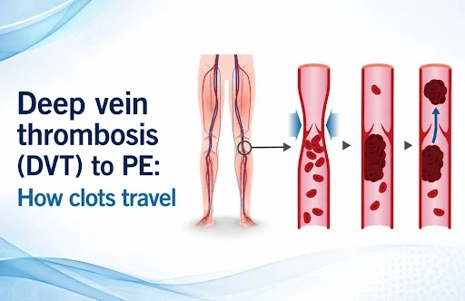
Deep vein thrombosis When someone develops sudden leg swelling, unexplained pain, or breathlessness that comes on quickly, the link between deep vein thrombosis (DVT) and pulmonary embolism (PE) becomes clinically significant. Knowing how a clot forms in the veins, breaks loose, and travels to the lungs gives doctors and medical students a clearer framework for diagnosis and treatment. This article walks through that pathway, the warning signs at each stage, how these conditions are diagnosed, and the core principles behind DVT treatment and pulmonary embolism treatment.

What Is Deep Vein Thrombosis?

DVT refers to a blood clot that forms inside a deep vein, most often in the leg  typically the calf, thigh, or pelvic veins. Depending on its size and location, the clot can partly or fully block normal blood flow through the vein.

Common signs include:

  • Swelling in one leg
  • Pain or tenderness in the affected limb
  • A feeling of warmth over the area
  • Redness or a change in skin color
  • Sometimes, no noticeable symptoms at all

That last point matters clinically  DVT doesn’t always announce itself. In some cases, a clot stays silent until it embolizes and causes problems elsewhere.

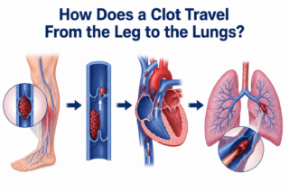
How Does a Clot Travel From the Leg to the Lungs?
Deep vein thrombosis

A Deep vein  thrombosis becomes a pulmonary embolism when a piece of the clot breaks free and travels through the bloodstream.

The general route looks like this:

Deep leg veins → inferior vena cava → right atrium → right ventricle → pulmonary arteries → lungs

Once a fragment of the clot detaches, it’s called an embolus. It moves through the venous system toward the right side of the heart and from there into the pulmonary circulation. If it becomes lodged in a pulmonary artery, that’s a pulmonary embolism.

How serious the resulting PE is depends largely on the embolus’s size, where it lodges, and how much cardiopulmonary reserve the patient has. A large clot can meaningfully disrupt blood flow through the lungs and put real strain on the right side of the heart.

Also read: Deep vein thrombosis treatment | Cardiologist in Hyderabad 

Why Early DVT Treatment Matters

Once DVT is diagnosed or strongly suspected, the treatment goal is to stop the clot from growing and to lower the risk that a piece of it breaks away.

Anticoagulant medication is the backbone of DVT treatment. These drugs don’t dissolve the existing clot directly, but by limiting further clot formation, they give the body time to break down the thrombus on its own.

The specific anticoagulation plan, which drug and for how long,, is tailored to the individual, taking into account:

  • Where the clot is and how extensive it is
  • The patient’s bleeding risk
  • Any history of prior DVT or PE
  • Whether cancer or another underlying condition is driving clot formation
  • Recent surgery, hospital stays, or long periods of immobility
  • Other relevant health conditions

In other words, treatment isn’t one-size-fits-all just because a clot is present; it has to match the patient’s broader clinical picture.

Recognizing When Deep vein thrombosis Has Become PE

Pulmonary embolism should be on the radar any time a patient with known or suspected DVT suddenly develops heart or lung symptoms.

Typical warning signs of PE include:

  • Sudden or unexplained shortness of breath
  • Chest pain that worsens with deep breaths
  • A racing or irregular heartbeat
  • Persistent cough
  • Coughing up blood
  • Dizziness or fainting

None of these symptoms is unique to PE on its own, which is exactly why proper clinical evaluation and testing are essential rather than guesswork.

A typical scenario: someone recovering from a long period of immobility first notices swelling in one calf. If they then develop sudden breathlessness and sharp chest pain that worsens with breathing, that combination should prompt urgent evaluation for DVT-related PE.

How Pulmonary Embolism Is Treated

The right approach to pulmonary embolism treatment depends on how sick the patient is, how much of the pulmonary circulation is blocked, and the risk that their condition could worsen.

Anticoagulation remains the mainstay for preventing further clotting. For patients who are hemodynamically unstable or have a more severe PE, more aggressive interventions may be warranted depending on the clinical picture and the resources available.

Diagnostic workup commonly includes:

  • D-dimer testing in the right clinical context
  • Compression ultrasound to check for DVT
  • CT pulmonary angiography to confirm PE
  • ECG and cardiac biomarkers to assess risk
  • Echocardiography in select cases, to check how the right side of the heart is coping

The goal isn’t just to find the clot  it’s to understand how dangerous it is and how urgently the patient needs treatment.

When to Involve a Pulmonary Embolism Specialist

A pulmonary embolism specialist becomes especially relevant in complex cases: an extensive PE, significant strain on the heart and lungs, or a situation where advanced procedures like catheter-based clot removal are being considered.

For patients or referring doctors searching for a top pulmonary embolism doctor, a pulmonary embolism specialist in India, or a pulmonary embolism doctor nearby, it’s worth prioritizing a clinician experienced specifically in venous thromboembolic disease, including catheter-based and multidisciplinary treatment options where they apply.

For clinicians, the key takeaway is that DVT and PE aren’t two unrelated diagnoses; they’re two stages of the same underlying disease process, venous thromboembolism, and should be assessed together rather than in isolation.

The DVT-to-PE Pathway, Step by Step

  1. A clot forms in a deep vein.
  2. Part of the clot breaks loose.
  3. The embolus travels through the venous system.
  4. It reaches the right side of the heart.
  5. It passes into the pulmonary arteries.
  6. It may block blood flow through the lungs.
  7. The result is a pulmonary embolism.

This chain of events is exactly why catching DVT early and treating it properly matters so much in preventing a potentially life-threatening complication.

Also read : Pulmonary Embolism in India: Emergency & Finding Specialist 

When to Seek Specialist Care Urgently

Sudden breathlessness, unexplained chest pain, coughing up blood, fainting, or a noticeably fast heart rate in someone with known or suspected DVT calls for immediate medical attention. PE can be life-threatening, and it’s not something to wait out or try to manage without professional evaluation.

For patients dealing with complex DVT or pulmonary embolism, Dr. C. Raghu offers specialized cardiovascular care, including catheter-based treatment for venous thrombosis and pulmonary vascular disease.

If you or someone you know has suspected or confirmed DVT or pulmonary embolism, consult Dr. C. Raghu for a specialist evaluation and a treatment plan suited to your specific condition.

pulmonary-embolism-in-india.webp

June 1, 2026 AbhijitHearty Life

A Clot That Travels to the Lung  and the Urgency It Demands

Pulmonary embolism (PE) is a sudden blockage of one or more pulmonary arteries, the blood vessels carrying blood from the heart to the lungs, by a clot that has usually traveled from the deep veins of the legs or pelvis. It is a potentially catastrophic emergency. A massive pulmonary embolism, in which a large clot obstructs the main pulmonary arteries, can produce acute right heart failure, hemodynamic collapse, and cardiac arrest within minutes. Even smaller emboli, if not recognized and treated, can be fatal.

In India, pulmonary embolism is substantially underdiagnosed. Limited awareness of its symptoms among patients and families, incomplete recognition among some clinicians, and the incorrect perception that PE is primarily a Western disease all contribute to diagnostic delays that cost lives. Improving awareness of pulmonary embolism — who is at risk, what it feels like, and what treatment options are available from a qualified pulmonary embolism specialist in India  is therefore genuinely life-saving.

Deep Vein Thrombosis and Pulmonary Embolism: The Same Condition, Different Stages

DVT treatment and pulmonary embolism management are two aspects of managing a single disease: venous thromboembolism (VTE). Deep vein thrombosis (DVT) is the formation of a blood clot in a deep vein, most commonly the deep veins of the calf, thigh, or pelvis. When a piece of that clot breaks off and travels through the venous system to the right heart and into the pulmonary arteries, pulmonary embolism results.

DVT classically presents with unilateral leg swelling, pain, tenderness, and redness, though a substantial proportion of DVT cases are asymptomatic and discovered only when PE occurs. Any asymmetric leg swelling in a patient with risk factors deserves urgent evaluation. Doppler ultrasound of the leg veins, combined with D-dimer blood testing, is the standard diagnostic pathway.

The risk factors for venous thromboembolism in India’s patient population are highly prevalent: prolonged immobility (hospitalization, long-haul flights, bed rest), recent major surgery (particularly hip and knee replacement), active cancer, pregnancy, oral contraceptive or hormone therapy use, obesity, and inherited thrombophilia (blood clotting disorders). Many patients who develop PE have two or more of these factors coexisting.


Read more: DVT to Pulmonary Embolism: The Risks and Prevention 

Recognising Pulmonary Embolism: Symptoms That Demand Immediate Action

Pulmonary embolism symptoms vary with the size of the embolism and the patient’s underlying cardiopulmonary reserve:

Sudden breathlessness is  often the most prominent symptom, appearing abruptly and without obvious cause. Any unexplained acute onset of breathlessness, particularly in a patient with PE risk factors, must be evaluated urgently for pulmonary embolism.

Pleuritic chest pain is  sharp chest pain that worsens with breathing or coughing, resulting from inflammation of the pleura (lung lining) adjacent to the embolic event.

Rapid heart rate (tachycardia) — nearly universal in significant PE, reflecting the cardiovascular stress of reduced pulmonary circulation.

Hemoptysis,  coughing blood-tinged sputum, occurs in pulmonary infarction when a segment of lung tissue loses blood supply.

Dizziness, presyncope, or syncope are  symptoms of hemodynamic compromise, indicating significant right heart strain.

In massive PE, a large central clot causes hemodynamic collapse the presentation is dramatic: severe breathlessness, hypotension, pallor, cyanosis, and potentially cardiac arrest. This constitutes an extreme cardiac emergency requiring immediate transfer to a hospital with pulmonary embolism treatment capabilities and, in appropriate cases, emergency mechanical intervention.

Any combination of sudden unexplained breathlessness, tachycardia, and recent leg swelling should trigger immediate medical evaluation. Delay is dangerous.

Read more: Dr. C. Raghu is one of the best doctors for TAVI/TAVR in Hyderabad  

Diagnosing Pulmonary Embolism

Diagnostic probability is first assessed clinically; validated scoring tools like the Wells PE Score quantify the pre-test probability of PE based on symptoms, signs, and risk factors. In low- to intermediate-probability patients, a D-dimer blood test can safely exclude PE when negative. In high-probability patients, imaging proceeds directly.

CT pulmonary angiography (CTPA) is the gold standard investigation; it demonstrates clots directly in the pulmonary arteries, defines the anatomical extent of embolic burden, and characterizes right heart strain on the same scan. Echocardiography provides rapid bedside assessment of right ventricular function in hemodynamically unstable patients who cannot be safely transported for CTPA.

Treatment: Stratified by Severity

Low-risk PE  normal blood pressure, and no right heart strain  is treated with anticoagulation. Direct oral anticoagulants (DOACs), particularly rivaroxaban and apixaban, are now first-line treatment, enabling outpatient or brief inpatient management for most low-risk cases.

Submassive PE with  normal blood pressure but echocardiographic or CT evidence of right ventricular dysfunction requires close monitoring and anticoagulation, with selective use of catheter-directed therapy for patients who deteriorate or are at high risk of clinical decompensation.

Massive PE  hemodynamic compromise constitutes an emergency requiring immediate restoration of pulmonary blood flow. Systemic thrombolysis (intravenous clot-dissolving drugs) is the primary intervention when not contraindicated. When systemic thrombolysis is contraindicated by bleeding risk or has failed to produce clinical improvement, catheter-based mechanical thrombectomy physical removal or fragmentation of a clot from the pulmonary arteries—is the advanced intervention that can be life-saving.


Read more: Hyderabad Cardiologist Reveals 5 Silent Heart Warning Signs  

Mechanical Thrombectomy: Advanced PE Care Available in India

Mechanical thrombectomy for pulmonary embolism involves navigating a catheter through the femoral vein, through the right heart, and into the pulmonary arteries, where dedicated devices aspirate or fragment the clot and restore blood flow without the systemic bleeding risk of intravenous thrombolytics. Large-bore aspiration thrombectomy systems and ultrasound-facilitated catheter-directed thrombolysis are the principal modalities used.

This advanced intervention requires specific training, a specialized catheterization laboratory team, and the rapid mobilization of a multidisciplinary PE Response Team (PERT). Dr. Raghu’s practice in Hyderabad maintains this PE treatment capability, offering patients with high-risk PE access to the most advanced catheter-based interventional options available.

For PE evaluation, DVT treatment, or urgent vascular consultation, contact Dr. Raghu’s team. For active emergencies, contact emergency services immediately.
Call Us: +91 95424 75650
Write to us: secretary@drraghu.com


Dr raghu logo


Dr raghu logo


Call us now if you are in a medical emergency need, we will reply swiftly and provide you with a medical aid.


For Appointment with Dr Raghu: +91 80085 36699
Dr Raghu’s Patient Coordinator: +91 72860 10203
Dr Raghu’s Office Experience: +91 90001 65962
Dr Raghu’s Social Media Communication Channel: +91 97046 51708

Dr. Raghu | Heart Specialist in Hyderabad
Yashoda Hospitals, Raj Bhavan Road, Somajiguda, 1st floor, Room No. 115, Hyderabad -500082, Telangana

Best Cardiologists in Hyderabad


© 2024, Dr. Raghu. All rights reserved. Design & Developed by AMSDigital.in

[gtranslate]
+91-9542475650