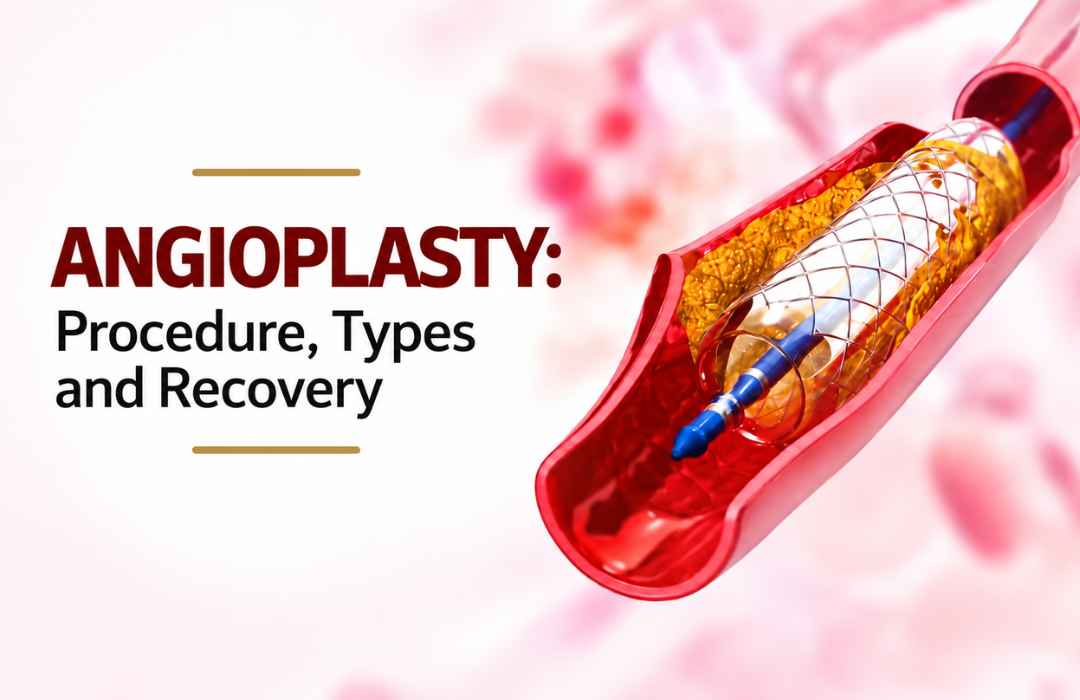
Angioplasty-Procedure-Types-and-Recovery.jpg

When a coronary artery is completely blocked, patients often want to know whether it can be reopened, what the procedure involves, and how long recovery may take. CTO Angioplasty in Heart treatment is a specialized form of coronary intervention used in selected patients with a chronic total occlusion. This guide explains what a CTO is, the main procedure approaches, who may be considered for treatment, recovery after CTO angioplasty, and why assessment by a Coronary Artery Disease Specialist matters.

What Is CTO Angioplasty?

CTO stands for chronic total occlusion. It refers to a coronary artery that has been completely blocked for a prolonged period. Over time, the body may develop smaller alternative blood vessels called collateral vessels that can supply some blood to the heart muscle beyond the blockage.

A CTO is different from a partially narrowed coronary artery. The blockage can contain hardened plaque and fibrous tissue, making it more technically challenging for a catheter and guidewire to cross.

CTO angioplasty, also called CTO PCI, is a catheter-based procedure that attempts to restore blood flow through the chronically blocked coronary artery. Whether it is appropriate depends on the patient’s symptoms, coronary anatomy, heart function, evidence of ischaemia or viable heart muscle, other coronary blockages, and overall health.

Current chronic coronary syndrome guidance emphasizes individualized assessment and shared decision-making when selecting a revascularization strategy.

Who May Be Considered for CTO Angioplasty?

Having a CTO does not automatically mean that angioplasty is required. Some patients may have adequate blood supply through collateral vessels and few or no symptoms, while others may have persistent symptoms or evidence that the blocked artery is affecting the heart muscle.

A Coronary Artery Disease Specialist may consider CTO PCI when factors such as the following are present:

  • Persistent or recurrent angina despite appropriate medical treatment
    Reduced exercise tolerance or symptoms affecting daily activities
    Evidence of significant ischaemia related to the territory supplied by the blocked artery
    Viable heart muscle that may benefit from improved blood flow
    Coronary anatomy that is suitable for PCI
    A clinical situation in which the expected benefits of revascularization outweigh its risks

For some patients, medication and lifestyle management may remain appropriate. In others, PCI or coronary artery bypass surgery may be considered. The decision is based on the individual rather than the blockage alone.

Types of CTO Angioplasty Procedures

The approach used during CTO PCI depends heavily on the location, length and characteristics of the blockage and the surrounding coronary anatomy.

Antegrade Approach

In an antegrade approach, the guidewire is directed toward the CTO from the usual starting point of the coronary artery.

This may be considered when the anatomy provides a suitable path through the occluded segment. The operator uses coronary imaging and procedural techniques to navigate the wire toward the distal part of the artery.

Retrograde Approach

A retrograde approach reaches the blockage from the opposite side of the occlusion. In selected cases, the guidewire can be directed through suitable collateral vessels to approach the distal portion of the CTO.

This can be useful when an antegrade strategy is difficult or does not provide a suitable route. It requires careful assessment of the coronary anatomy and specialized procedural expertise.

Hybrid CTO PCI

A hybrid approach uses more than one CTO PCI strategy depending on what happens during the procedure.

For example, the operator may begin with an antegrade strategy and change to another technique if the initial pathway proves unsuitable. This flexibility allows the procedure to be adapted to the patient’s anatomy rather than forcing every CTO into the same treatment method.

How Is CTO Angioplasty Performed?

Although CTO PCI is more complex than many routine angioplasty procedures, the overall process follows familiar catheter-based principles.

The procedure may involve:

  • Detailed assessment: Previous angiograms and other cardiac imaging are reviewed to understand the blockage and surrounding vessels.
    Arterial access: A catheter is introduced through an appropriate artery, commonly through the wrist or groin.
    Guidewire navigation: Specialized guidewires are carefully directed toward and, when possible, across the chronic blockage.
    Balloon treatment: Once an appropriate pathway has been established, balloon angioplasty can be used to widen the treated segment.
    Stent placement: A coronary stent may be placed when appropriate to help maintain the opened vessel.
    Final assessment: The result is checked before the catheters are removed and the patient moves to post-procedure monitoring.


The American Heart Association describes PCI as a catheter-based treatment that can open narrowed or blocked coronary artery segments, with patients monitored after catheterization for bleeding, blood pressure, heart rhythm and other concerns.

CTO Angioplasty vs Bypass Surgery

CTO angioplasty and coronary artery bypass grafting (CABG) are different ways of restoring blood flow to the heart.

CTO PCI is performed through catheters and generally does not require an open surgical incision. CABG uses blood vessels from another part of the body to create a new route around blocked coronary arteries.

Neither option is automatically appropriate for every patient. The choice may depend on:

  • Number and location of blocked arteries
    Complexity of coronary disease
    Heart function
    Diabetes and other medical conditions
    Surgical risk
    Coronary anatomy
    Previous procedures
    Patient preferences and expected outcomes

For complex coronary disease, a cardiology team may compare PCI, surgery and medical treatment before recommending a strategy. The 2024 ESC guidance specifically emphasizes selecting the revascularization approach according to the patient’s clinical profile, coronary anatomy, procedural factors and preferences.

CTO Angioplasty Recovery: What to Expect

Recovery after CTO angioplasty varies. A straightforward catheter-based procedure may allow relatively rapid return to normal activities, but CTO PCI can require more extensive procedural planning and monitoring than routine PCI.

First 24 Hours

Immediately after the procedure, the medical team monitors the patient’s heart rhythm, blood pressure and catheter access site. The access area is checked for bleeding, swelling or other complications.

First Few Days

The patient is generally advised to rest and gradually resume normal activities according to the treating team’s instructions. Care of the wrist or groin access site is particularly important.

Following Weeks

Activity can usually increase progressively when medically appropriate. Some patients may also be advised to participate in cardiac rehabilitation, depending on their overall cardiac condition and treatment plan.

Follow-up appointments are important for reviewing symptoms, medications, risk factors and overall cardiovascular health.

Medicines After CTO Angioplasty

Medication is an important part of care after coronary intervention, particularly when a stent has been implanted.

Depending on the patient’s circumstances, prescribed treatment may include:

  • Antiplatelet medication
    Cholesterol-lowering medication
    Blood-pressure medication
    Anti-anginal treatment
    Other medicines related to diabetes or cardiovascular risk


Patients should not stop or change prescribed heart medication without speaking with their treating clinician. Current ESC guidance includes antiplatelet therapy as an important component of treatment after PCI, with the regimen and duration individualized according to clinical and bleeding risk.

What Are the Risks of CTO Angioplasty?

CTO PCI is technically demanding and carries risks that need to be discussed before treatment. Potential complications can include:

  • Bleeding or bruising at the catheter access site
    Blood-vessel injury
    Coronary artery perforation
    Abnormal heart rhythms
    Heart attack
    Contrast-related complications
    Need for additional intervention or, rarely, emergency surgery
    Re-narrowing or repeat blockage


The exact risk depends on the patient’s health, coronary anatomy and procedural complexity. This is why careful pre-procedure planning and appropriate specialist assessment are important.

When Should a Patient Contact a Doctor After CTO PCI?

After cardiac catheterization or angioplasty, new or worsening symptoms should not be ignored.

Patients should seek medical advice for concerns such as:

  • New or worsening chest pain
    Shortness of breath
    Persistent or increasing bleeding from the access site
    Significant swelling or pain around the access site
    Dizziness or fainting
    Fever
    A very fast, very slow or irregular heartbeat


The American Heart Association advises patients to report chest pain, shortness of breath, significant access-site swelling or bleeding and other concerning symptoms after cardiac catheterization.

If severe chest discomfort, breathing difficulty or other possible heart-attack symptoms occur, emergency medical care should be sought immediately rather than waiting for a routine appointment.

Choosing a Specialist for CTO Care

Because CTO PCI can be more technically complex than routine angioplasty, patients may want to ask about the specialist’s experience with chronic total occlusions and the range of treatment options available.

Useful questions include:

  • Is the blockage suitable for CTO PCI?
    What approach is being considered?
    Are there alternative treatment options?
    Would bypass surgery or medical management be more appropriate?
    What complications are relevant to the patient’s anatomy?
    What follow-up will be required after treatment?


Patients seeking a Top Cardiologist in Hyderabad can review the available cardiac services and arrange a specialist consultation through Dr. C. Raghu’s website.

Related Cardiac Conditions and Specialist Care

Some patients undergoing coronary evaluation may also have other cardiovascular conditions. For example, older adults may have coronary artery disease alongside aortic valve disease, while unexplained breathlessness may require assessment for several possible causes.

For patients with significant aortic stenosis, a TAVI Expert in Hyderabad may evaluate whether transcatheter aortic valve replacement is appropriate. Similarly, patients with suspected pulmonary embolism require a separate diagnostic pathway and, when confirmed, appropriate Pulmonary Embolism Treatment in Hyderabad.

These conditions should not be assumed to be related to a CTO. Each requires appropriate clinical evaluation.

Also read this : What is coronary angioplasty? Everything you need to know

Moving Forward After a CTO Diagnosis

A chronic total occlusion can sound alarming, but the diagnosis does not by itself determine the treatment. Some patients may be managed with medication and risk-factor control, while selected patients may benefit from CTO PCI or another revascularization strategy.

For someone experiencing persistent chest discomfort, reduced exercise tolerance or other concerning cardiac symptoms, the practical next step is a detailed evaluation rather than delaying care or assuming that every complete blockage requires the same procedure.

Patients in Hyderabad, Telangana, Chennai and elsewhere in India can learn more about CTO Angioplasty in Heart and coronary artery disease care by visiting Dr. C. Raghu’s website and arranging an appropriate specialist consultation. The goal is to understand the anatomy, available options and expected recovery clearly so that treatment decisions can be made with the treating cardiac team.

Find how an CTO Angioplasty in Heart in Hyderabad can help improve your heart health and quality of life. Consult Dr. C. Raghu, an experienced interventional cardiologist and TAVI Expert in Hyderabad, specializing in structural heart disease at Yashoda Hospital. Schedule your consultation for personalized cardiac care.
deep-vein-thrombosis.webp

September 4, 2026 AbhijitHearty Life

Deep vein thrombosis When someone develops sudden leg swelling, unexplained pain, or breathlessness that comes on quickly, the link between deep vein thrombosis (DVT) and pulmonary embolism (PE) becomes clinically significant. Knowing how a clot forms in the veins, breaks loose, and travels to the lungs gives doctors and medical students a clearer framework for diagnosis and treatment. This article walks through that pathway, the warning signs at each stage, how these conditions are diagnosed, and the core principles behind DVT treatment and pulmonary embolism treatment.

What Is Deep Vein Thrombosis?

DVT refers to a blood clot that forms inside a deep vein, most often in the leg  typically the calf, thigh, or pelvic veins. Depending on its size and location, the clot can partly or fully block normal blood flow through the vein.

Common signs include:

  • Swelling in one leg
  • Pain or tenderness in the affected limb
  • A feeling of warmth over the area
  • Redness or a change in skin color
  • Sometimes, no noticeable symptoms at all

That last point matters clinically  DVT doesn’t always announce itself. In some cases, a clot stays silent until it embolizes and causes problems elsewhere.

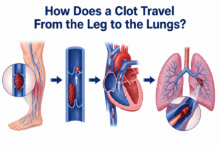
How Does a Clot Travel From the Leg to the Lungs?
Deep vein thrombosis

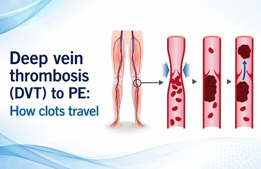
A Deep vein  thrombosis becomes a pulmonary embolism when a piece of the clot breaks free and travels through the bloodstream.

The general route looks like this:

Deep leg veins → inferior vena cava → right atrium → right ventricle → pulmonary arteries → lungs

Once a fragment of the clot detaches, it’s called an embolus. It moves through the venous system toward the right side of the heart and from there into the pulmonary circulation. If it becomes lodged in a pulmonary artery, that’s a pulmonary embolism.

How serious the resulting PE is depends largely on the embolus’s size, where it lodges, and how much cardiopulmonary reserve the patient has. A large clot can meaningfully disrupt blood flow through the lungs and put real strain on the right side of the heart.

Also read: Deep vein thrombosis treatment | Cardiologist in Hyderabad 

Why Early DVT Treatment Matters

Once DVT is diagnosed or strongly suspected, the treatment goal is to stop the clot from growing and to lower the risk that a piece of it breaks away.

Anticoagulant medication is the backbone of DVT treatment. These drugs don’t dissolve the existing clot directly, but by limiting further clot formation, they give the body time to break down the thrombus on its own.

The specific anticoagulation plan, which drug and for how long,, is tailored to the individual, taking into account:

  • Where the clot is and how extensive it is
  • The patient’s bleeding risk
  • Any history of prior DVT or PE
  • Whether cancer or another underlying condition is driving clot formation
  • Recent surgery, hospital stays, or long periods of immobility
  • Other relevant health conditions

In other words, treatment isn’t one-size-fits-all just because a clot is present; it has to match the patient’s broader clinical picture.

Recognizing When Deep vein thrombosis Has Become PE

Pulmonary embolism should be on the radar any time a patient with known or suspected DVT suddenly develops heart or lung symptoms.

Typical warning signs of PE include:

  • Sudden or unexplained shortness of breath
  • Chest pain that worsens with deep breaths
  • A racing or irregular heartbeat
  • Persistent cough
  • Coughing up blood
  • Dizziness or fainting

None of these symptoms is unique to PE on its own, which is exactly why proper clinical evaluation and testing are essential rather than guesswork.

A typical scenario: someone recovering from a long period of immobility first notices swelling in one calf. If they then develop sudden breathlessness and sharp chest pain that worsens with breathing, that combination should prompt urgent evaluation for DVT-related PE.

How Pulmonary Embolism Is Treated

The right approach to pulmonary embolism treatment depends on how sick the patient is, how much of the pulmonary circulation is blocked, and the risk that their condition could worsen.

Anticoagulation remains the mainstay for preventing further clotting. For patients who are hemodynamically unstable or have a more severe PE, more aggressive interventions may be warranted depending on the clinical picture and the resources available.

Diagnostic workup commonly includes:

  • D-dimer testing in the right clinical context
  • Compression ultrasound to check for DVT
  • CT pulmonary angiography to confirm PE
  • ECG and cardiac biomarkers to assess risk
  • Echocardiography in select cases, to check how the right side of the heart is coping

The goal isn’t just to find the clot  it’s to understand how dangerous it is and how urgently the patient needs treatment.

When to Involve a Pulmonary Embolism Specialist

A pulmonary embolism specialist becomes especially relevant in complex cases: an extensive PE, significant strain on the heart and lungs, or a situation where advanced procedures like catheter-based clot removal are being considered.

For patients or referring doctors searching for a top pulmonary embolism doctor, a pulmonary embolism specialist in India, or a pulmonary embolism doctor nearby, it’s worth prioritizing a clinician experienced specifically in venous thromboembolic disease, including catheter-based and multidisciplinary treatment options where they apply.

For clinicians, the key takeaway is that DVT and PE aren’t two unrelated diagnoses; they’re two stages of the same underlying disease process, venous thromboembolism, and should be assessed together rather than in isolation.

The DVT-to-PE Pathway, Step by Step

  1. A clot forms in a deep vein.
  2. Part of the clot breaks loose.
  3. The embolus travels through the venous system.
  4. It reaches the right side of the heart.
  5. It passes into the pulmonary arteries.
  6. It may block blood flow through the lungs.
  7. The result is a pulmonary embolism.

This chain of events is exactly why catching DVT early and treating it properly matters so much in preventing a potentially life-threatening complication.

Also read : Pulmonary Embolism in India: Emergency & Finding Specialist 

When to Seek Specialist Care Urgently

Sudden breathlessness, unexplained chest pain, coughing up blood, fainting, or a noticeably fast heart rate in someone with known or suspected DVT calls for immediate medical attention. PE can be life-threatening, and it’s not something to wait out or try to manage without professional evaluation.

For patients dealing with complex DVT or pulmonary embolism, Dr. C. Raghu offers specialized cardiovascular care, including catheter-based treatment for venous thrombosis and pulmonary vascular disease.

If you or someone you know has suspected or confirmed DVT or pulmonary embolism, consult Dr. C. Raghu for a specialist evaluation and a treatment plan suited to your specific condition.


what-is-a-pulmonary-embolism.jpg

July 13, 2026 AbhijitHearty Life

Pulmonary embolism You’re sitting at work when suddenly you can’t catch your breath. Your chest feels tight. Your heart is racing. You feel like you’re going to pass out. You’re scared genuinely terrified.

Your coworker calls an ambulance. At the hospital, after tests, the doctor tells you, “You have a pulmonary embolism.” A blood clot has traveled to your lungs.

Your world stops. You’ve heard that phrase before “pulmonary embolism” and you know it’s serious. You need to understand what just happened, why it happened, and what comes next.

What Exactly Is a Pulmonary Embolism?

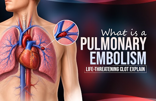
A pulmonary embolism—often called PE—occurs when a blood clot travels through your veins and lodges in the blood vessels of your lungs. This clot blocks blood flow, preventing oxygen from being transferred to your blood.

The clot usually starts in your legs or pelvis. It breaks loose and travels through your venous system, through your right heart, and into your lungs, where it gets stuck—too large to fit through the narrowing vessels.

Think of it like this: imagine a road with traffic flowing normally. Suddenly, a large object blocks the road. Traffic backs up. Things are chaos.That’s what happens in your lungs when a clot blocks blood flow.

Also read: DVT to Pulmonary Embolism: The Risks and Prevention  

Why You Get Blood Clots: Risk Factors

Several situations increase your clot risk:

Immobility: Sitting for long periods—long flights, road trips, bed rest after surgery, and office work. When you don’t move, blood pools in your legs instead of circulating normally. Pooled blood clots.

Surgery: After surgery, blood clotting mechanisms activate. You’re immobile during recovery. You have trauma to your tissues. This combination creates perfect conditions for clots.

Cancer: Cancer cells activate clotting mechanisms. Cancer patients have high PE risk.

Birth control pills or hormone therapy: Estrogen increases clotting tendency.

Pregnancy and postpartum period: Pregnancy naturally increases clotting tendency. After delivery, you’re at even higher risk.

Previous blood clots: If you’ve had one clot, you’re at higher risk for another.

Genetic clotting disorders: Some people have genetic variations making their blood clot more easily.

Obesity: Extra weight increases clot risk.

Immobilized leg: Fractures, casts, or reduced mobility in one leg increase risk.

Recent hospitalization: Being bedridden increases risk.

Smoking: Smoking damages blood vessels and increases clotting.

Heart disease: Certain heart conditions increase PE risk.

Symptoms: The Warning Signs

Pulmonary embolism symptoms vary based on the clot’s size and location. Small clots might cause mild symptoms. Large clots can cause sudden collapse.

Common symptoms include:

  • Sudden severe shortness of breath
  • Chest pain, especially when breathing deeply
  • Rapid heartbeat
  • Dizziness or lightheadedness
  • Fainting
  • Anxiety or sense of impending doom
  • Cough (sometimes with blood-tinged sputum)
  • Leg pain or swelling (if clot originated there)

Important: These symptoms warrant emergency evaluation. Don’t wait. Call 911 or go to the emergency room immediately.

Why PE is So Serious

Your lungs’ job is to oxygenate blood. A clot blocking lung blood vessels prevents oxygenation.

Your oxygen levels drop. Your brain, heart, and other organs aren’t getting sufficient oxygen.

Your right heart overworks. The right ventricle has to pump harder against the blocked vessels. It can become exhausted.

Your blood pressure drops. If the clot is large, your cardiovascular system can collapse.

You go into shock. Organ systems fail without sufficient oxygen.

This is why pulmonary embolism is a medical emergency. It can be fatal. Sudden massive PE can cause sudden death.

Diagnosis: How Doctors Identify PE

If your doctor suspects PE, several tests confirm the diagnosis:

CT Pulmonary Angiography (CTPA): This is usually the first test. A CT scanner with special imaging shows blood clots in your lungs’ vessels. It’s quick, accurate, and readily available.

D-dimer blood test: This blood test measures a substance released when clots break down. If D-dimer is normal, PE is very unlikely. If elevated, further testing is needed.

Chest X-ray: Might show pneumonia or other lung problems, but usually looks normal in PE.

Echocardiogram: Ultrasound of your heart. Shows if your right heart is strained.

Arterial blood gas: Measures oxygen and carbon dioxide levels in your blood.

Ventilation-perfusion scan: Shows areas of your lungs receiving ventilation (air) but not perfusion (blood flow). Mismatches suggest PE.

Also Read: Atrial Fibrillation treatment in India: Complete Guide 

Pulmonary Embolism Treatment: Your Options

Once PE is diagnosed, immediate treatment begins.

Anticoagulation Therapy: Blood thinners prevent clots from growing and new clots from forming. They don’t dissolve existing clots—your body does that naturally—but they prevent expansion while your body works to break down the clot.

Medications include:

  • Heparin: Given intravenously in the hospital
  • Low molecular weight heparin: Injected subcutaneously, can be outpatient
  • Warfarin: Oral blood thinner for long-term management
  • Direct oral anticoagulants: Newer medications like apixaban or rivaroxaban

Thrombolytic Therapy (Clot-Busting): For massive PEs or hemodynamically unstable patients, medications that dissolve clots might be used. These medications have risks but can be lifesaving in severe PE.

Mechanical Thrombectomy: For very large clots or patients who can’t tolerate blood thinners, a pulmonary embolism specialist might perform mechanical thrombectomy. A catheter-based device removes clot material directly.

IVC Filter: In rare cases where you can’t take blood thinners, an inferior vena cava (IVC) filter might be placed. This filter catches clots traveling from legs before they reach your lungs.

Recovery and Aftercare

After initial PE treatment:

You’ll stay in the hospital for a few days to ensure stability and adjust medications.

You’ll take blood thinners for months to years, depending on circumstances.

Regular blood tests monitor your blood’s clotting tendency.

Follow-up imaging might be done to ensure clots are resolving.

You’ll gradually resume activity as you recover.

Most people recover completely from their first PE if treated promptly.

Post-PE Complications

Some people develop complications after PE:

Post-thrombotic syndrome: Chronic leg swelling and pain from valve damage in leg veins.

Chronic thromboembolic pulmonary hypertension (CTEP): Some clots don’t fully dissolve, leading to permanent lung scarring and heart strain.

Recurrent PE: Some people have another PE despite treatment.

This is why finding the right pulmonary embolism doctor and following their guidance completely is critical.

Finding Your PE Specialist

You need a pulmonary embolism specialist in India or your local area who:

  • Understands PE completely
  • Can manage your acute care
  • Monitors for complications
  • Helps you navigate blood thinner therapy
  • Works with you long-term

A pulmonary embolism specialist in Hyderabad can provide comprehensive care if you’re in that region.

For complicated cases or patients not responding well, the best doctor for pulmonary embolism at a major medical center might be appropriate.

Prevention: Reducing Your Risk

If you’re at risk for PE:

Move regularly. On flights or road trips, get up and walk every 2 hours.

Compression socks. These prevent blood pooling in your legs during immobility.

Leg exercises. Flex your calves while sitting to activate leg muscles and promote blood flow.

Hydration. Dehydration increases clot risk. Drink plenty of water.

Blood thinner prophylaxis. If you’re having surgery or hospitalization, your doctor might prescribe preventive blood thinners.

Stop smoking. Smoking damages blood vessels.

Manage your weight. Obesity increases PE risk.

Control medical conditions. Manage heart disease, cancer risk factors, and other conditions that increase clot risk.

Life After Pulmonary Embolism

Most people recover completely from PE. You’ll likely take blood thinners for several months to several years, depending on whether this was provoked (caused by a temporary situation) or unprovoked (no clear cause).

You’ll have restrictions initially bu gradually return to normal activities.

You’ll attend follow-up appointments and take medications as prescribed.

You’ll be aware of PE symptoms and seek immediate care if symptoms recur.

Many people go on to live normal, full lives after PE. The key is treating it promptly and following your doctor’s guidance carefully.

Moving Forward

Pulmonary embolism is serious. It demands immediate medical attention. It requires ongoing management. But it’s treatable, and most people recover well.

If you’ve experienced PE or are at high risk, understand your risk factors. Modify them where possible. Know the warning signs. Seek immediate care if symptoms develop.

Work with qualified specialists. Take your medications. Attend your follow-ups. Be an active participant in your care.

Your life after PE can be full and normal. It requires attention to your health, but it’s absolutely possible.

If you’ve experienced a pulmonary embolism or are concerned about your risk, expert medical guidance is essential. Consult with Dr. C. Raghu, a cardiologist in Hyderabad specializing in cardiovascular emergencies and blood clot management.


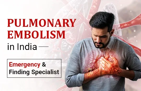
pulmonary-embolism-in-india.webp

June 1, 2026 AbhijitHearty Life

A Clot That Travels to the Lung  and the Urgency It Demands

Pulmonary embolism (PE) is a sudden blockage of one or more pulmonary arteries, the blood vessels carrying blood from the heart to the lungs, by a clot that has usually traveled from the deep veins of the legs or pelvis. It is a potentially catastrophic emergency. A massive pulmonary embolism, in which a large clot obstructs the main pulmonary arteries, can produce acute right heart failure, hemodynamic collapse, and cardiac arrest within minutes. Even smaller emboli, if not recognized and treated, can be fatal.

In India, pulmonary embolism is substantially underdiagnosed. Limited awareness of its symptoms among patients and families, incomplete recognition among some clinicians, and the incorrect perception that PE is primarily a Western disease all contribute to diagnostic delays that cost lives. Improving awareness of pulmonary embolism — who is at risk, what it feels like, and what treatment options are available from a qualified pulmonary embolism specialist in India  is therefore genuinely life-saving.

Deep Vein Thrombosis and Pulmonary Embolism: The Same Condition, Different Stages

DVT treatment and pulmonary embolism management are two aspects of managing a single disease: venous thromboembolism (VTE). Deep vein thrombosis (DVT) is the formation of a blood clot in a deep vein, most commonly the deep veins of the calf, thigh, or pelvis. When a piece of that clot breaks off and travels through the venous system to the right heart and into the pulmonary arteries, pulmonary embolism results.

DVT classically presents with unilateral leg swelling, pain, tenderness, and redness, though a substantial proportion of DVT cases are asymptomatic and discovered only when PE occurs. Any asymmetric leg swelling in a patient with risk factors deserves urgent evaluation. Doppler ultrasound of the leg veins, combined with D-dimer blood testing, is the standard diagnostic pathway.

The risk factors for venous thromboembolism in India’s patient population are highly prevalent: prolonged immobility (hospitalization, long-haul flights, bed rest), recent major surgery (particularly hip and knee replacement), active cancer, pregnancy, oral contraceptive or hormone therapy use, obesity, and inherited thrombophilia (blood clotting disorders). Many patients who develop PE have two or more of these factors coexisting.


Read more: DVT to Pulmonary Embolism: The Risks and Prevention 

Recognising Pulmonary Embolism: Symptoms That Demand Immediate Action

Pulmonary embolism symptoms vary with the size of the embolism and the patient’s underlying cardiopulmonary reserve:

Sudden breathlessness is  often the most prominent symptom, appearing abruptly and without obvious cause. Any unexplained acute onset of breathlessness, particularly in a patient with PE risk factors, must be evaluated urgently for pulmonary embolism.

Pleuritic chest pain is  sharp chest pain that worsens with breathing or coughing, resulting from inflammation of the pleura (lung lining) adjacent to the embolic event.

Rapid heart rate (tachycardia) — nearly universal in significant PE, reflecting the cardiovascular stress of reduced pulmonary circulation.

Hemoptysis,  coughing blood-tinged sputum, occurs in pulmonary infarction when a segment of lung tissue loses blood supply.

Dizziness, presyncope, or syncope are  symptoms of hemodynamic compromise, indicating significant right heart strain.

In massive PE, a large central clot causes hemodynamic collapse the presentation is dramatic: severe breathlessness, hypotension, pallor, cyanosis, and potentially cardiac arrest. This constitutes an extreme cardiac emergency requiring immediate transfer to a hospital with pulmonary embolism treatment capabilities and, in appropriate cases, emergency mechanical intervention.

Any combination of sudden unexplained breathlessness, tachycardia, and recent leg swelling should trigger immediate medical evaluation. Delay is dangerous.

Read more: Dr. C. Raghu is one of the best doctors for TAVI/TAVR in Hyderabad  

Diagnosing Pulmonary Embolism

Diagnostic probability is first assessed clinically; validated scoring tools like the Wells PE Score quantify the pre-test probability of PE based on symptoms, signs, and risk factors. In low- to intermediate-probability patients, a D-dimer blood test can safely exclude PE when negative. In high-probability patients, imaging proceeds directly.

CT pulmonary angiography (CTPA) is the gold standard investigation; it demonstrates clots directly in the pulmonary arteries, defines the anatomical extent of embolic burden, and characterizes right heart strain on the same scan. Echocardiography provides rapid bedside assessment of right ventricular function in hemodynamically unstable patients who cannot be safely transported for CTPA.

Treatment: Stratified by Severity

Low-risk PE  normal blood pressure, and no right heart strain  is treated with anticoagulation. Direct oral anticoagulants (DOACs), particularly rivaroxaban and apixaban, are now first-line treatment, enabling outpatient or brief inpatient management for most low-risk cases.

Submassive PE with  normal blood pressure but echocardiographic or CT evidence of right ventricular dysfunction requires close monitoring and anticoagulation, with selective use of catheter-directed therapy for patients who deteriorate or are at high risk of clinical decompensation.

Massive PE  hemodynamic compromise constitutes an emergency requiring immediate restoration of pulmonary blood flow. Systemic thrombolysis (intravenous clot-dissolving drugs) is the primary intervention when not contraindicated. When systemic thrombolysis is contraindicated by bleeding risk or has failed to produce clinical improvement, catheter-based mechanical thrombectomy physical removal or fragmentation of a clot from the pulmonary arteries—is the advanced intervention that can be life-saving.


Read more: Hyderabad Cardiologist Reveals 5 Silent Heart Warning Signs  

Mechanical Thrombectomy: Advanced PE Care Available in India

Mechanical thrombectomy for pulmonary embolism involves navigating a catheter through the femoral vein, through the right heart, and into the pulmonary arteries, where dedicated devices aspirate or fragment the clot and restore blood flow without the systemic bleeding risk of intravenous thrombolytics. Large-bore aspiration thrombectomy systems and ultrasound-facilitated catheter-directed thrombolysis are the principal modalities used.

This advanced intervention requires specific training, a specialized catheterization laboratory team, and the rapid mobilization of a multidisciplinary PE Response Team (PERT). Dr. Raghu’s practice in Hyderabad maintains this PE treatment capability, offering patients with high-risk PE access to the most advanced catheter-based interventional options available.

For PE evaluation, DVT treatment, or urgent vascular consultation, contact Dr. Raghu’s team. For active emergencies, contact emergency services immediately.
Call Us: +91 95424 75650
Write to us: secretary@drraghu.com


pulmonary-embolism.webp

January 20, 2026 AbhijitHearty Life

People get really worried about blood clots and the problems they can cause. For instance, if you do not treat venous thrombosis,s it can become a very serious disease called pulmonary embolism at some point. This blog is about the signs of embolism, the things that put you at risk of getting pulmonary embolism, or Pulmonary Embolism Treatment,what you can do to prevent pulmonary embolism in India, and the symptoms of pulmonary embolism you should know about before you go see a pulmonary embolism specialist or a doctor who treats acute pulmonary embolism. They can help you get away and make sure you get the care you need to deal with pulmonary embolism.

Key Takeaways

Deep vein thrombosis, which is what DVT Treatment is, happens when a blood clot forms in a vein, usually in the leg. This blood clot in the vein can be very bad news. Sometimes the clot in the vein can break loose and move to the lungs. When the clot from the vein goes to the lung,s it can cause a big problem called Pulmonary Embolism. This Pulmonic Embolism is very serious. Can be life-threatening. So when someone has a Pulmonic Embolism from a deep vein thrombosis, they need to see a pulmonary embolism specialist. The specialist for embolism needs to act fast to help the person with the deep vein thrombosis and the Pulmonic Embolism.

When you are not moving around for a time or you have had recent surgeries or you are overweight or obese or you smoke or you are pregnant or you are older than 60 years or you have underlying medical conditions like cancer and heart problems, these things can increase the chance of a blood clot forming. The risk factors I mentioned like being overweight or obese, smoking and having underlying conditions like cancer and heart problems can cause a clot to form, and this can lead to a Pulmonary Embolism. Pulmonary Embolism is a condition that can happen because of these risk factors, including being immobile for a long time, recent surgeries and other things, like cancer and heart problems.

If you have Deep Vein Thrombosis, you might notice that one of your legs is swollen, it hurts and it looks red. It feels warm. On the other hand if you have the best doctor for pulmonary embolism, you will feel like you cannot get enough air and you will have a sudden pain in your chest that gets worse when you take a deep breath or cough. Sometimes you might even cough up blood. Your heart will beat really fast. If any of these things happen to you you should go to the emergency room away or find a Pulmonary Embolism doctor near me.

You can also read this : Deep vein thrombosis treatment | Cardiologist in Hyderabad

To stop Deep Vein Thrombosis from happening, you need to do things. You have to exercise and drink plenty of water.

Deep Vein Thrombosis can be prevented by not sitting for long.

You should also wear socks called compression stockings.

You have to take medicine that helps prevent blood clots, which is given by a doctor who treats lung problems in India, a pulmonary embolism specialist, in India.

In essence, it means saving lives through early detection. Therefore, such symptoms and risks should be noted to assist patients in getting early treatment in the hands of an expert pulmonary embolism doctor.

Awareness of Risk Factors Associated with DVT and Pulmonary Embolism

DVT and Pulmonary Embolism risk factors are health states, life habits, and medical histories that combine and result in the arrival of a clot.

What DVT and Pulmonary Embolism are all about:

Deep vein thrombosis occurs in a deep vein, most commonly within a leg, when a blood clot forms. That clot can free itself when onboard a new trip to the lungs, and that is where the medical emergency, a pulmonary embolism, is created, needing a pulmonary embolism specialist to fall under his therapy.

DVT includes a swollen leg, pain, and red patches that may occur. However, for Pulmonary Embolism, signs may include sudden breathlessness or chest pain or coughing up blood. Early diagnosis by a pulmonary embolism doctor is critical as this might avert severe complications.

DVT and Pulmonary Embolism Symptoms

The swelling, pain, and warmth often seen in one leg are symptoms of DVT. On the other hand, symptoms of pulmonary embolism tend to be much more severe, including sudden shortness of breath, sharp chest pains, rapid heartbeat, or coughing blood. Deaths happen as well from misidentifying patients in healthcare facilities. Immediate evaluation by a pulmonary embolism doctor near me can save lives.

Causes of DVT and Pulmonary Embolism

The cause of thrombus formation is immobility, surgery, or underlying medical conditions that slow the blood flow enough for it to occur. However, in pregnant women and certain cancers, high estrogen levels can increase risk for thrombus formation, leading toa condition that travels to the pulmonary arteries, where it needs to be urgently treated by a pulmonary embolism specialist in India.

At risk for DVT and Pulmonary Embolism

This includes a clotting disorder, prolonged immobility, obesity, smoking habits, and older age. Other possible factors are pregnancy and heart disease. Knowledge of these risk factors makes one take the initiative to seek early evaluation from the best doctor for pulmonary embolism.

Connection Between DVT and Pulmonary Embolism

DVT is, therefore, within deep veins; that is, within those associated veins, clots will form. When any clot that is formed into the veins of the legs dislodged and breaks into circulation, it will eventually clog somewhere in the lungs. Hence, one reason that makes this important connection to early treatment and preventive care for this is that it benefits from being seen by a pulmonary embolism specialist doctor.

Prevention of DVT and Pulmonary Embolism: Strategic Approaches

Risk-lowering lifestyle changes:

Regular exercise, hydration, and a balanced diet, while avoiding prolonged sitting can help improve circulation and reduce the risk of forming a Pulmonary Embolism. Simple daily habits may be done to reduce and even champion vein protection.

Interventions to Prevent DVT and Pulmonary Embolism

Patients will be required for early intervention; this may involve prescribing anticoagulants and using compression stockings to minimize the risk of clot formation.

Detect and Treat Early

Early diagnosis is the key to successful management in most cases; failure to diagnose early may lead to complications and even death. Timely intervention by the best doctor of pulmonary embolism will, therefore, do a lot in saving the life of an individual to severe long-term injuries.

Educate Yourself about DVT and PE

That way, when DVT Treatment  and Pulmonary Embolism strike, people will be able to know it in advance and alert those individuals most likely to be a sufferer among them. The symptoms of DVT and PE , and the risk factors and preventive measures for DVT and PE will always keep everyone alert to promptly contact a pulmonary embolism specialist in India whenever the situation demands it.

You can also read this : Chronic Thromboembolic Pulmonary Hypertension

Conclusion

DVT and PE are serious conditions, and these may also be preventable. Hence, being aware of the signs, the risk factors, and how to prevent the occurrence of such incidents is vital. Early diagnosis and management of a patient by a pulmonary embolism doctor or specialist doctor of pulmonary embolism would greatly reduce the risks of complications and result in better outcomes.

If you or a loved one notice symptoms of DVT or pulmonary embolism, don’t delay—consult Dr. C. Raghu, an experienced specialist for Pulmonary Embolism treatment in India, for timely diagnosis, advanced care, and life-saving intervention. Get direction


Pulmonary-Embolism-Treatment.webp

June 19, 2025 AbhijitHearty Life

Pulmonary embolism or deep vein thrombosis is a serious medical condition. Blood clot formation is its primary cause. When this clot enters the bloodstream, there's a high risk for it to reach the lungs. Consequently, the clot hinders the lung arteries and curtails the oxygen and blood flow. Usually, it develops inside the deeper veins in the legs. When detected, the pulmonary embolism treatment must be immediate as it may prove to be fatal without immediate care.


Dr raghu logo


Dr raghu logo


Call us now if you are in a medical emergency need, we will reply swiftly and provide you with a medical aid.


For Appointment with Dr Raghu: +91 80085 36699
Dr Raghu’s Patient Coordinator: +91 72860 10203
Dr Raghu’s Office Experience: +91 90001 65962
Dr Raghu’s Social Media Communication Channel: +91 97046 51708

Dr. Raghu | Heart Specialist in Hyderabad
Yashoda Hospitals, Raj Bhavan Road, Somajiguda, 1st floor, Room No. 115, Hyderabad -500082, Telangana

Best Cardiologists in Hyderabad


© 2024, Dr. Raghu. All rights reserved. Design & Developed by AMSDigital.in

[gtranslate]
+91-9542475650