cci-coronary_angioplasty.jpg


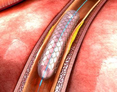
Coronary arteries are the blood vessels that delivers oxygenated blood to the heart muscle. But, sometimes these blood vessels may get narrowed or blocked due to accumulation of cholesterol plaque. To open the blockage and restore the function of arteries, coronary angioplasty is performed.

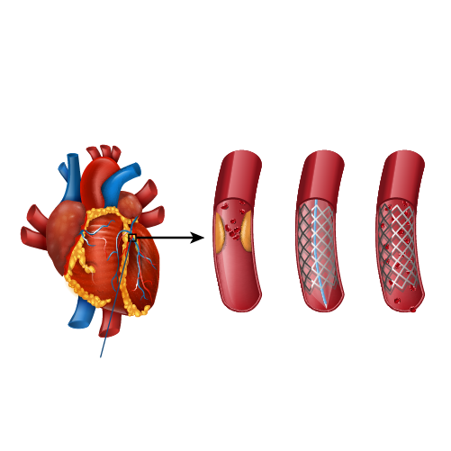
A coronary angioplasty involves the temporary insertion of a tiny balloon inside the artery to open the blockage. It is usually combined with the placement of a stent (a metal mesh tube) to widen the artery and prevent the further chance of narrowing.
coronary angioplasty

Why is coronary angioplasty done ?

Coronary angioplasty is done to treat narrowing or blockage of the heart’s blood vessels:

  • during or after a heart attack.
  • Blood vessel narrowing leading to poor heart function.
  • For relief of chest pain (angina) due to reduced blood supply to heart.

Risks of coronary angioplasty

Usually, it is a safe procedure, but like all other procedures, it also carries certain risks like:

Common Issues/Risks after angiography
Bruising Common to have bruise at groin or arm Lasts for few weeks Not to be worried about
Allergic reactions Usually related to contrast medicine used History of previous allergy – higher risk Present as rash and headache
Bleeding at the site of entry (Hematoma) Usually subsides in a few days But in case swelling is increasing and painful contact your physician immediately


Serious complications after coronary angiography Less than 1 in 1000 chance People with severe heart disease are at high risk Discuss with Cardiologist before procedure
Heart attack A serious medical emergency where the heart’s blood supply is suddenly blocked
Stroke A serious medical condition that occurs when the blood supply to the brain is interrupted
Loss of blood supply to limb Damage to the artery in the arm or groin in which the catheter was inserted, with the blood supply to the limb possibly being affected
Kidney injury Damage to the kidneys caused by the contrast dye
Radiation injury Tissue damage caused by X-ray radiation if the procedure is prolonged

What happens before the procedure?

A physical examination and blood test are done to evaluate the overall health condition. The person needs to follow instructions provided before undergoing the procedure:

1. Inform the doctor about:

  • Any allergies
  • The use of current medicines, including vitamin and mineral supplements
  • Any blood disorder
  • Any surgery you may had
  • The past and present medical condition

2. Medicines:

Tell the doctor if you are using blood thinners or any anti-diabetic medicines. The doctor will either advise you to stop or change the dose of the medication.

3. Food and fluid restrictions:

  • Avoid heavy meals, such as meat, fried or fatty foods eight hours before the procedure. Take light foods such as toast and cereal.
  • Fast for six hours before the procedure.
  • Stay hydrated, drink plenty of fluid up to two hours before the procedure.

In most of the cases, you will be discharged on the same day of the hospital, so ask someone to accompany you to the hospital.

What happens during the procedure?

Local anaesthesia would be administered to numb the area where the catheter would be inserted. Mostly, a catheter is inserted through the wrist, and sometimes through the groin. Under fluoroscopy (a type of X-ray), the catheter will be guided to the diseased artery. Then a special medicine (called contrast or dye) will be injected into the blood vessels supplying the heart. This will delineate the presence of obstruction to blood flow. 
Then a wire is passed across the obstruction and a balloon is inflated at the site of obstruction to compress the obstructing plaque in the artery. The balloon expansion will compress the plaques into the wall of the blood vessel and improve the blood flow. Once the blood flow is improved, the catheter will be removed, and a stent would be placed to prevent the risk of further blockage. Finally, the catheter will be removed and the puncture site is sealed.

What to expect after the procedure?

The nurse will check the vitals of the patient. Depending on the condition, the doctor would advise you the hospital stay. You will be advised not to bend or cross the leg for a few days if the procedure is performed through the groin region. Additionally, some other tests like X-rays and an electrocardiogram (ECG) would be done to check the condition. Before discharge, the doctor will give you certain instructions regarding wound care, medications and lifestyle.

Recovery after coronary angioplasty

Recover quickly and keep your heart healthy by following these tips:

  • Quit smoking.
  • Limit the intake of alcohol.
  • Take the prescribed medicines.
  • Check the cholesterol levels regularly.
  • Maintain a healthy body weight.
  • Have a healthy and well-balanced diet.
  • Exercise regularly.

Know your numbers

It’s important to periodically monitor and maintain the following numbers to prevent future cardiac events

  • LDL cholesterol – less than 55 mg/dL
  • Blood pressure – less than 120/80 mm Hg
  • Blood sugar – HbA1C less than 7.0%

Book Online Consultaion


    Download: Coronary Angioplasty Blog
    GET DOWNLOAD PDF NOW

    Subscribe the Hearty Life Blogs


      disease-condtions-deep_vein_thrombosis.png

      When a blood clot (thrombus) is formed in one or more of the deep veins of your body (usually in the legs), it is known as deep vein thrombosis (DVT). Sometimes this clot becomes detached from the inner walls of the blood vessel and travels to another part inside the body. DVT may develop due to an underlying medical condition particularly the one which affects the clotting of the blood.

      Causes and risk factors:

      It can be caused by the factors which prevent the normal circulation of the blood and it’s clotting, such as long periods of rest (during a particular medical condition), injury to the vein, surgery and certain medications.

      The risk factors are as follows:

      • Inherited blood clotting disorder
      • Prolonged bed rest
      • Surgery
      • Pregnancy
      • Contraceptives or hormone replacement therapy
      • Obesity
      • Smoking
      • Cancer
      • Heart failure
      • Inflammatory bowel disease
      • Age (usually older than 60 years but can also occur at any age)

      Complications:

      The following are the complications which may arise due to DVT:

      Pulmonary embolism: In Pulmonary embolism, a thrombus present in another part of your body usually a leg, travels to the blood vessel of the lung and blocks the blood circulation, leading to a life-threatening condition.

      Post-phlebetic syndrome (post thrombotic syndrome): set of signs and symptoms seen after a blood clot has formed.

      Symptoms:

      The person may be asymptomatic during the formation of the thrombus, however, during post thrombotic period when it has caused damage to the veins, the blood flow to the affected body part may be compromised. The following symptoms are observed:

      • Persistent swelling of the leg or arm (edema)
      • Leg pain or tenderness while standing
      • Skin discoloration in the affected arm or leg
      • Veins in the arm or leg appearing larger than normal

      Sometimes the development of a serious health complication such as pulmonary embolism is responsible for the symptoms in the DVT patients which are as follows:

      • Shortness of breath
      • Chest pain
      • Cough (blood may be coughed out as well)
      • Back pain
      • Profuse sweating
      • Lightheadedness
      • Blue nails or lips

      These symptoms appear suddenly, and an immediate medical attention is required.

      Diagnosis:

      The diagnosis is initiated by a thorough clinical examination and involves review of the patient’s medical history. Based upon these findings, the physician will categorize the patient into low or high risk DVT. Further tests may be considered to rule out other health problems and to confirm the diagnosis, which are as follows:

      • Blood tests: These are considered to evaluate any blood disorders that increase risk of DVT.
      • D- dimer test: In this test, the levels of a protein called D-dimer is assessed. In patients with DVT, high levels of this protein can be found in the blood.
      • Duplex ultrasonography: It is a non-invasive evaluation of the blood flow in the arteries and the veins of a patient. But this test is not preferred for a thrombus located very deep inside the body such as pelvis.
      • Venography: It is a specialized type of an x-ray procedure wherein a dye is injected into a vein of the foot, and the blood clot if present is detected.
      • Magnetic resonance imaging (MRI): It is performed in patients for whom ultrasound examination is inappropriate or unfeasible. It can even detect a thrombus present in the patient’s pelvis and the thigh.

      Prevention:

      A recent World Thrombosis Day Ipsos survey showed that there is less awareness of DVT in countries around the world. Thus, the first step in prevention is making the public aware of DVT. The preventive measures are different depending on the person’s condition who are at risk of developing DVT:

      During pregnancy

      In pregnant women, certain medication may be prescribed if she has risk factors such as likelihood of having a cesarean birth, bed rest, history of DVT, and inherited thrombophilia.

      During travel or in longer hours of sitting posture:

      • Drink lots of fluids.
      • Wear loose fitting clothes.
      • Walk and stretch at regular intervals.
      • Use special stockings to improve blood flow and to prevent blood clots but consult your health care provider before using it.

      Treatment:

      Treatment for DVT includes certain medicines and therapies, which are as follows:

      Medications:

      • Anticoagulants: They prevent further formation of blood clots but don’t dissolve the already existing ones. Most common anticoagulants used are heparin and warfarin. Warfarin is not recommended during pregnancy.
      • Thrombin inhibitors: These are recommended for people who can’t take heparin.
      • Thrombolytic medicines: These are recommended only during life threatening condition.

      Compression stockings:

      It may be recommended to prevent swelling and to avoid post-thrombotic symptoms in the patients.

      Surgical treatment:

      Surgical treatment may be performed when medications fail to dissolve the clots.

      Book Online Consultaion


        Deep Vein Thrombosis Blog

        DOWNLOAD PDF

        Subscribe the Hearty Life Blogs


          disease-condtions-thromboembolism.jpg

          Thromboembolism refers to a condition where a blood clot formed in the blood vessel unplugs itself and flows in the blood stream to block another blood vessel leading to obstruction of blood flow. Although clotting is a normal bodily function, if a clot occurs where it is not required, it may lead to serious problems.

          Types of Thromboembolism:

          The type of thromboembolism differs depending on the area in which the clot may appear. It can obstruct the blood flow in the veins, arteries, brain, gastrointestinal tract or kidneys.

          Thromboembolism can be further classified as below:

          Venous Thromboembolism:

          A venous thromboembolism is a thrombus (blood clot) formed in a vein. The blood flow in the veins is slow when compared to arteries which can increase the likelihood of the blood clots forming in these vessels.

          It can be categorized into three different types

          • Superficial Vein Thrombosis
            The clot may occur in a superficial vein, close to the surface of the body. Usually, it is not serious until it enters the deep veins.
          • Deep Vein Thrombosis (DVT)
            Deep vein thrombosis occurs within the deep veins, and mostly occurs within the legs.
          • Pulmonary embolism (PE)
            Pulmonary embolism is a medical emergency in which the thrombus blocks the blood vessel in the lungs.

          Renal Vein Thrombosis (RVT)

          A renal vein thrombosis is a thrombosis that occurs in the veins that drain blood away from the kidneys. These clots reduce the ability of the kidneys to clean and filter the blood.

          Arterial Thrombosis (atherothrombosis)

          Arterial thrombosis is much less common than venous thrombosis. It can have similar risks. Usually arterial thrombosis may lead to necrosis of the tissue.

          A thromboembolism in the coronary artery can cause a heart attack. If blood supply to the brain is disrupted, the patient may suffer a stroke.

          Symptoms:

          Signs and symptoms of venous thromboembolism include the following

          DVT

          • Pain in arm or leg, usually in the thigh or calf
          • Swollen leg or arm
          • Reddening or warming of the skin
          • Red streaks on the skin

          PE

          • Shortness of breath
          • Chest pain under your rib cage that can get worse when you take a deep breath
          • Rapid heart rate
          • Feeling lightheaded or passing out
          • Accentuated second heart sound

          Arterial Thrombosis

          • Muscle pain or spasm in the affected area
          • Numbness and tingling in the arm or leg
          • Pale color of the arm or leg (pallor)
          • Weakness of an arm or leg

          If the condition is severe, it may lead to blister formation and shedding of the skin, leading to tissue necrosis.

          Symptoms of a clot in an organ vary with the organ involved but may include:

          • Pain in the part of the body that is involved
          • Temporary decrease in organ function

          Causes:

          The blood clots can occur due to injury to a vein, consequence of a surgery, use of certain medications and lack of movement of the limbs.

          In the case of PE, the blood clot may block the blood vessels of the lungs. The affected portion of the lung may die due to loss of blood supply, the condition is called as pulmonary infraction, making it difficult to provide oxygen to the rest of the body.

          Occasionally, the blood vessels can be blocked by substances other than blood like collagen, part of a tumour, air bubbles etc.

          Risk factors:

          The common risk factors include

          • Inheriting a blood-clotting disorder
          • Prolonged immobility which may be due to bed rest or long trips
          • Injury or surgery
          • Pregnancy
          • Birth control pills or hormone replacement therapy
          • Being overweight or obese
          • Smoking
          • Cancer
          • Heart failure
          • Inflammatory bowel disease
          • Family history of deep vein thrombosis or pulmonary embolism

          Diagnosis:

          The diagnosis of thromboembolism includes the following tests which could be considered depending on the type:

          D-dimer: The blood sample is tested for the presence of D-dimer which is a marker for the presence of blood clots.

          Duplex Ultrasound: This is an imaging test in which the presence of clots is identified using ultrasound waves.

          Pulse oximetry: In this test, a sensor attached on the end of the finger of the patient helps to measure the level of oxygen in the blood.

          Arterial blood gas: The blood drawn from the artery is checked for oxygen levels present in it.

          Chest X-ray: This test may not be useful in finding the presence of clots, but can help to rule out a clot.

          Other diagnostic tests may include ELISA, pulmonary angiography, venography, echocardiography, helical computed tomography of pulmonary vessels etc.

          Treatment:

          Treatment of thromboembolism includes:

          Blood thinners: These are anticoagulant drugs which prevent formation of new clots while the body works to break up the previous clots present. They include heparin, low-molecular-weight heparin, apixaban, edoxaban, rivaroxaban, and warfarin.

          Clot-busting drugs: These are intravenous injections to dissolve the clots in the case of life-threatening situations. They include drugs belonging to the class of tissue plasminogen activators.

          Surgical and other procedures:

          • Removal of clot: Surgery is considered only if the condition is life threatening. The clot is removed using a catheter threaded through the blood vessel.
          • Vein filter: A catheter can also be used to position a filter in the inferior vena cava which helps to stop the clots from being carried into the lungs.

          Prevention:

          If the patient is at a risk of the condition, the following preventive measures may be considered to reduce the occurrence:

          • Use of blood thinners
          • Use of compression stockings
          • Use of intermittent pneumatic compression devices that automatically squeeze the legs to keep blood flowing

          Other preventive measures include

          • Regular exercise
          • Maintaining healthy weight
          • Cessation of smoking

          Consideration during travel include

          • Walk every 1 to 2 hours
          • Stretch legs and move around on the seat
          • Drink lots of fluids
          • Avoid smoking and consumption of alcohol before journey

          Book Online Consultaion


            Thromboembolism Blog

            DOWNLOAD PDF

            Subscribe the Hearty Life Blogs


              Patent-Ductus-Arteriosus-1-1200x688.webp

              Patent ductus arteriosus (PDA) is a congenital heart condition in which there is an opening between the pulmonary artery and the aorta. The ductus arteriosus is a small connection in the fetal heart helps oxygen rich blood to by-pass the immature baby’s lungs and flow into the body.


              Dr raghu logo


              Dr raghu logo


              Call us now if you are in a medical emergency need, we will reply swiftly and provide you with a medical aid.


              For Appointment with Dr Raghu: +91 80085 36699
              Dr Raghu’s Patient Coordinator: +91 72860 10203
              Dr Raghu’s Office Experience: +91 90001 65962
              Dr Raghu’s Social Media Communication Channel: +91 97046 51708

              Dr. Raghu | Heart Specialist in Hyderabad
              Yashoda Hospitals, Raj Bhavan Road, Somajiguda, 1st floor, Room No. 115, Hyderabad -500082, Telangana

              Best Cardiologists in Hyderabad


              © 2024, Dr. Raghu. All rights reserved. Design & Developed by AMSDigital.in

              [gtranslate]
              +91-9542475650