What is an Atrial Septal Defect or ASD?
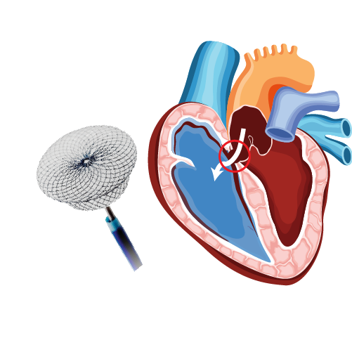
Heart consists of four chambers, of which upper two chambers are called as atria and lower two chambers are called as ventricles. Atrial septal defect (ASD) is a type of congenital heart defect, in which there is an abnormal opening or a hole in interatrial septum (dividing wall between two atria). This opening allows the passage of pulmonary venous blood from left atrium to right atrium, causing mixing of oxygenated and deoxygenated blood in right atrium and increasing the flow of blood to lungs. The increased blood flow may increase the workload of the lungs, and eventually cause damage to heart and lungs.
What causes an ASD?
The exact cause of ASD remains unclear. However, it is believed that during fetal developmental stages, a hole is present in the interatrial septum, which gradually closes before birth or during infancy. If the hole persists, it is called an atrial septal defect.
Types of Atrial Septal Defect or ASD?
Based on the location and development of ASD, it is classified into four types:
- Ostium secundum ASD: It occurs in the middle part of the interatrial septum. This is the most common type of ASD and accounts for 75% of all atrial septal defects. This type of ASD is commonly detected in adults in their third and fourth decades of life. Some can be detected in children when an abnormal heart sound is detected at the time of a routine health check or vaccination visit.
- Ostium primum ASD (20%): It occurs in the lower part of the interatrial septum, adjacent to atrioventricular (AV) valves. It usually occurs as a part of other congenital heart defects. This defect is usually detected in early life as this is associated with many complications.
- Sinus venosis ASD (4%): It occurs in the upper part of the interatrial septum, near the veins that drain into the right and left atrium. This is usually identified in third and fourth decade adults.
- Coronary sinus ASD (<1%): It occurs in the interatrial septum between the coronary sinus and the left atrium. This is very uncommon and patients are asymptomatic.
Signs and symptoms
Usually after birth, babies who have ASD may not show any associated signs and symptoms. But, symptoms may appear during adulthood around the age of 30 years. Most of them don’t have any symptoms even after many years.
Some of the common symptoms associated with ASD are:
- Heart murmur, a swishing sound that can be heard through a stethoscope
- Heart palpitations
- Arrhythmias (abnormal heart rhythms)
- Fatigue (feeling tired mainly after exercise)
- Shortness of breath
- Swelling of legs, feet or abdomen
- Stroke
- Lung infections such as pneumonia
Risk factors:
Genetics and environmental factors usually play a role in congenital heart defects. Some conditions during pregnancy may confer high risk for heart defects,
Complications of an untreated ASD:
- Right-sided heart failure
- Arrhythmias
- Increased risk of a stroke
- Shortened life span
- Pulmonary hypertension (increased blood flow to the lungs results in increased blood pressure in the lung arteries)
- Eisenmenger syndrome (pulmonary hypertension causing permanent lung damage)
Diagnosis:
Most ASDs are diagnosed incidentally during regular heath check-ups. If a heart murmur is heard during auscultation, you may be advised to undergo further tests to confirm the diagnosis of ASD:
- Echocardiogram: It is a specific diagnostic test for ASD, and provides a detailed image of the heart and blood flow through its chambers. The echocardiogram may show the blood flow through the interatrial septum and the size of defect in the septum.
- Chest X-ray: It helps to identify enlarged heart and lung changes.
- Electrocardiogram (ECG): It involves recording the electrical activity of the heart, to help identify arrhythmias.
- Cardiac catheterization: A thin, flexible tube called catheter is inserted into the blood vessel at the groin or arm and is guided to the heart. This test is helpful to determine the function of heart and its valves, and to measure the blood pressure in the lungs.
- Magnetic resonance imaging (MRI): It uses magnetic and radio waves to create three dimensional images of the heart and other organs. This test is recommended if the ASD is not clearly diagnosed with echocardiogram.
- Computerized tomography (CT) scan: It uses a series of X-rays to create detailed images of heart; mainly used if echocardiogram doesn’t help to diagnose ASD clearly.
Treatment:
Treatment of ASD depends on the age at diagnosis and the size, location and severity of the defect. Small ASDs might not need any treatment because it closes on its own. The doctor may recommend surgery if the atrial septal defect is large, even with fewer symptoms to prevent problems later in life. Treatment of ASD includes medications and surgery.
- Medications usually do not help in closing the hole, but reduces the signs and symptoms associated with ASD and risks of complications after surgery. Drugs such as beta blockers (to maintain regular heartbeat) and anticoagulants (to prevent the formation of blood clots) are used.
- Surgery includes cardiac catheterization and open-heart surgery.
Device closure of ASD
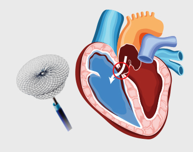
In cardiac catheterization, the doctor closes the hole in the septum with a mesh patch or a plug in the defect place using a catheter. The heart tissue then grows around the mesh slowly, sealing the hole permanently. This procedure is mainly performed to repair only the secundum type of atrial septal defects. Defects not amenable to closure with device may need open-heart surgery.
In open-heart surgery, the defect may be closed with stitches or a special patch. This procedure is done under general anesthesia, mainly for repairing primum, sinus venosus and coronary sinus atrial septal defects.
Prevention of ASD:
Atrial septal defects cannot be prevented, but following certain measures during pregnancy might be beneficial to prevent the risk of ASD, such as:
- Immunity test for rubella: If the person is not immune to rubella, it is necessary to get vaccinated.
- Monitoring current health conditions and medications use: Pre-existing health conditions and usage of any medications should be carefully monitored during pregnancy to prevent the risk of ASD.
- Reviewing family medical history: If a person has a family history of congenital defects, it is advised to visit a genetic counsellor before becoming pregnant to know the risks of atrial septal defects.
Our Specialities
- Conditions
- Acute limb ischemia
- Chronic limb ischemia
- Aortic stenosis
- Mitral valve stenosis
- Mitral valve regurgitation
- Atrial fibrillation
- Tachycardia
- Bradycardia
- Palpitations
- High blood pressure
- Atrial septal defect
- Ventricular septal defect
- Patent ductus arteriosus
- Cardiac amyloidosis
- Hypertrophic cardiomyopathy
- Varicose veins
- Deep vein thrombosis (DVT)
- Myocarditis
- Endocarditis
- Pericarditis
- Peripheral arterial disease
- Pulmonary artery hypertension
- Pulmonary embolism
- Cath lab procedures:
- Coronary Angiogram
- Primary Angioplasty
- Coronary Angioplasty
- CHIP Angioplasty
- Aortic valve replacement surgery
- Mitral valve replacement surgery
- Device closure for Atrial septal defect
- Device closure for Ventricular septal defect
- Device closure for Patent Ductus Arteriosus
- Transcatheter aortic valve replacement (TAVR)
- Inferior vena cava (IVC) filter
- LA appendage closure
- Fistuloplasty
- Balloon mitral valvotomy
- 24 hours emergency services
- Clinics- weekly basis/monthly basis/ Yearly basis
- Prevention of cardiovascular diseases
- Diagnosis
BOOK AN APPOINTMENT

DR. RAGHU
Cardiology Coronary, Vascular and
Structural Interventions
Conditions & Diseases
Angioplasty
Aortic Stenosis
Atrial Fibrillation
