Coronary-angioplasty-2.webp

July 1, 2026

Coronary angioplasty: Your chest feels tight. You’ve been getting shortness of breath lately, especially when you climb stairs or walk briskly. You’re worried. Your doctor has mentioned something called coronary angioplasty, but you don’t understand what it is or whether you really need it.

You’re not alone. Many middle-aged people are facing coronary artery disease without fully understanding their treatment options. The term “angioplasty” sounds intimidating. The word “stent” makes you nervous. But here’s the truth: understanding what these procedures are and how they can help you is empowering. It gives you control over your health decisions.

Understanding Coronary angioplasty Artery Disease
Coronary angioplasty

Before we discuss angioplasty, you need to understand the problem it solves.

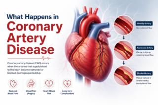
Your heart is a muscle that needs oxygen to work. Coronary arteries deliver oxygen-rich blood to your heart. These are the highways of your cardiovascular system. Over time, for various reasons—high cholesterol, high blood pressure, smoking, diabetes, and stress—fatty deposits build up inside these arteries. This buildup is called plaque.

Coronary artery disease happens when these arteries narrow. Blood flow decreases. Your heart struggles to get enough oxygen, especially during activity or stress. You feel chest pain. You feel exhausted. You feel vulnerable.

This is serious. Completely blocked arteries lead to heart attacks.

What Exactly is Coronary Angioplasty?

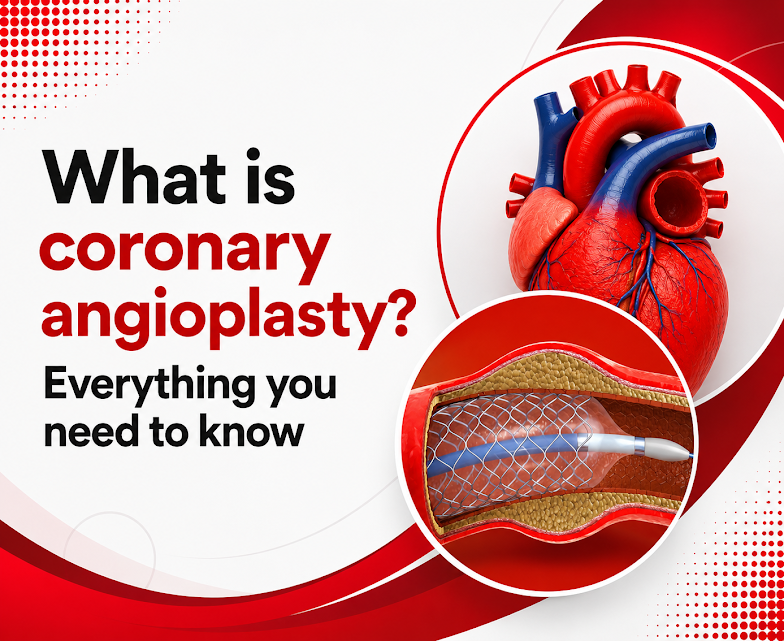
Let’s demystify this. Coronary angioplasty is a minimally invasive procedure that opens blocked or narrowed coronary arteries. It’s not surgery in the traditional sense—no large incisions, no general anesthesia, no weeks of recovery.

Here’s how it works:

Step One: Angiogram Your coronary artery disease doctor first performs an angiogram. A thin catheter (tube) is inserted through an artery in your groin or arm. This catheter is guided to your heart. A special dye is injected through the catheter, and X-rays are taken. These images show exactly where your arteries are blocked or narrowed. You’re awake during this procedure, though mildly sedated.

Step Two: Balloon Inflation Once the blocked area is identified, a balloon catheter is advanced to that location. When the catheter is in position, the balloon is inflated. This balloon pushes the plaque against the artery wall, widening the artery and restoring blood flow. You might feel slight chest discomfort—this is normal and temporary.

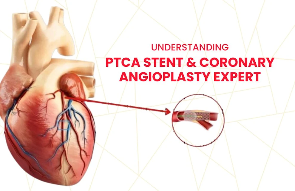
Step Three: Stent Placement In most modern angioplasty procedures, a stent is placed. Think of a stent as a tiny mesh tube. It keeps the artery open after the balloon is removed, preventing the artery from narrowing again. Most stents today are drug-eluting, meaning they release medication that prevents scar tissue buildup.

The entire procedure typically takes 30 minutes to an hour.

Why Your Doctor Recommends Angioplasty

Your coronary artery disease specialist recommends angioplasty for specific reasons:

  • You have significant blockages causing symptoms
  • You have blockages that could cause a heart attack
  • Medical management (medications) isn’t sufficient
  • You’ve had a recent heart attack
  • You’re having a heart attack right now

Not all coronary artery disease requires angioplasty. Some people do well with medications. But when angioplasty is appropriate, it can be life-changing.

Also read: Pulmonary Embolism in India: Emergency & Finding Specialist  

The Difference Between Angioplasty and Angiogram

Many people confuse these terms. Let me clarify:

An angiogram is a diagnostic test. It shows what’s happening in your arteries.

Angioplasty is a treatment. It opens blocked arteries. It often includes stent coronary placement.

You might have an angiogram and learn that you don’t need angioplasty. Or you might have an angiogram and immediately proceed to angioplasty during the same procedure.

Recovery and Aftercare coronary angioplasty

Here’s what’s wonderful about angioplasty: recovery is relatively quick.

After the procedure:

  • You’ll stay in the hospital overnight or possibly go home the same day
  • You’ll have a bandage over the catheter insertion site
  • You can resume light activities within a few days
  • You return to normal activities within 1-2 weeks
  • You’ll take medications to prevent clotting and manage your condition

Compared to open-heart surgery, this is minimal disruption to your life.

Types of Angioplasty

Standard Angioplasty (PTCA): Percutaneous Transluminal Coronary Angioplasty is the traditional form, often with stent placement (PTCA Stent). It works well for most blockages.

Specialized Procedures: For particularly challenging blockages, chip angioplasty (rotational atherectomy) might be used. This technique actually rotates a burr that smooths hardened plaque. It’s more specialized and requires experienced operators.

For chronic total occlusions arteries completely blocked specialized techniques might be employed.

Finding Your Specialist in Hyderabad

You need an experienced coronary artery disease doctor who performs angioplasty regularly. You need someone who has handled complications and knows how to navigate the most challenging cases.

The best angioplasty expert in India will have extensive training, excellent outcomes, and a compassionate approach to patient care. They’ll explain everything clearly, answer your questions, and work with you to determine whether angioplasty is truly the right choice for your situation.

In Hyderabad, you have access to heart specialists in Hyderabad who bring world-class expertise to this procedure.

Life After Angioplasty

You need to understand: angioplasty treats the blockage, but it doesn’t cure the underlying coronary artery disease. You’ll still need to:

  • Take medications as prescribed (usually including blood thinners, blood pressure medications, cholesterol medications)
  • Make lifestyle changes—diet, exercise, stress management
  • Stop smoking, if applicable
  • Control diabetes and blood pressure
  • Attend regular follow-ups

The good news? Most people do extremely well after angioplasty. Blood flow is restored. Symptoms improve dramatically. Quality of life increases significantly.

Possible Complications (Rare, But Real)

While generally safe, angioplasty does carry small risks:

  • Bleeding at the catheter site
  • Artery perforation
  • Allergic reaction to dye
  • Stent restenosis (renarrowing)
  • Blood clots

This is why you need an experienced operator and proper aftercare.

Also read: CHIP Angioplasty: Advanced Coronary Artery Disease Treatment Explained  

Prevention is still best.

Remember: the best treatment is prevention. If you don’t have coronary artery disease yet but are at risk, focus on:

  • Not smoking
  • Managing stress
  • Exercising regularly
  • Eating healthy
  • Maintaining healthy weight
  • Controlling blood pressure and cholesterol
  • Managing diabetes

Moving Forward coronary angioplasty Doctor

If you’re dealing with coronary artery disease, you’re not facing a death sentence. Modern interventions like angioplasty have revolutionized how we treat this condition. Many people go on to live full, active lives after successful angioplasty.

The key is working with experienced specialists who understand both the procedure and your individual circumstances.

Take the first step toward better heart health. Consult with Dr. C. Raghu, an experienced interventional cardiologist in Hyderabad specializing in coronary interventions. Schedule your appointment.
Call Us +91 95424 75650 
Abhijit

Coronary-Artery-Disease-in-India.jpg

May 15, 2026

Coronary artery disease (CAD) kills more Indians than any other single condition. It strikes earlier in the Indian population than in Western countries, often a decade sooner,  driven by a combination of genetic susceptibility to dyslipidemia and insulin resistance, high rates of diabetes, rising rates of obesity and physical inactivity, tobacco use, and a diet increasingly influenced by processed food.

Yet the treatment available today — when coronary artery disease is detected early enough and managed by a skilled coronary artery disease specialist — is remarkably effective. Coronary angioplasty in heart arteries, supported by modern drug-eluting stents and evidence-based medication, gives patients with even severe coronary disease a very real path back to normal life.

The barrier is not treatment availability. It is awareness: patients who do not know the symptoms, who dismiss warning signs, or who do not understand what a coronary angioplasty and angiogram involve delay seeking help in ways that cost them heart muscle, cardiac function, and, sometimes, their lives.

This guide walks every reader through the complete pathway from recognizing symptoms through the diagnostic angiogram to PTCA stent placement and recovery—because understanding this pathway is genuinely life-saving.

Also read: Understanding PTCA Stent & Coronary Angioplasty expert

What Happens in Coronary Artery Disease

coronary artery disease

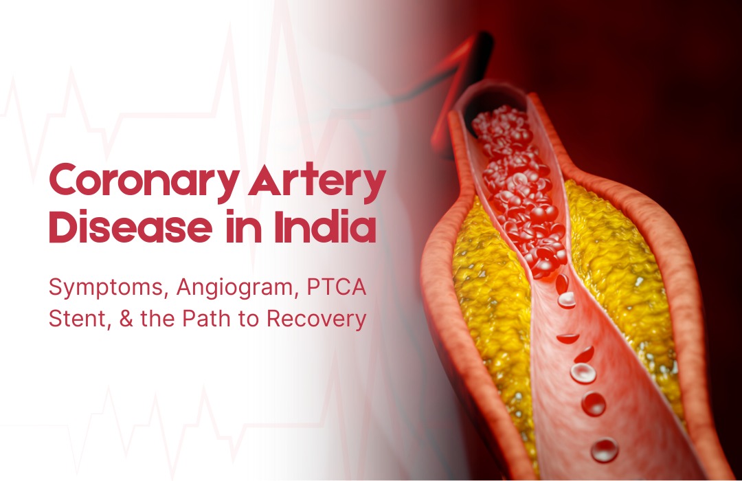
Coronary arteries are the three main blood vessels that supply oxygenated blood to the heart muscle itself. Coronary artery disease is the progressive accumulation of atherosclerotic plaque deposits of cholesterol, inflammatory cells, calcium, and connective tissue within the walls of these arteries. As plaques grow, they narrow the artery’s lumen, reducing the volume of blood the heart muscle receives.

In early disease, there are no symptoms. As narrowings become significant, the heart muscle receives adequate blood at rest but insufficient blood during exertion, producing the symptom of stable angina. When a plaque ruptures suddenly, as can happen without warning at any level of physical exertion, it triggers the formation of an acute blood clot that can completely block the artery, causing a heart attack 

Heart attacks cause permanent heart muscle damage in proportion to the time the artery remains blocked. Opening the blocked artery within 90 minutes of symptom onset with emergency coronary angioplasty (primary PCI) salvages the most muscle and produces the best survival outcomes. Every minute matters.

Recognising the Symptoms: What to Know, and What to Take Seriously

Stable angina is exertional chest discomfort—a tightness, pressure, heaviness, or aching in the center of the chest that appears predictably during physical activity (walking uphill, climbing stairs, carrying loads) and relieves with rest within a few minutes. It may radiate to the left arm, jaw, or back.

In the Indian population, and particularly in patients with diabetes, coronary artery disease sometimes presents atypically without classic chest pain,but with unexplained breathlessness on exertion, unusual fatigue, or a feeling of heaviness or indigestion. These symptoms in any patient with cardiovascular risk factors deserve a prompt cardiac evaluation, not reassurance.

Acute coronary syndromes,  unstable angina and heart attack, present with chest discomfort that is more severe and longer-lasting (more than 20 minutes) and may occur at rest. Associated symptoms, including sweating, nausea, breathlessness, or syncope, increase the likelihood of an acute event. These symptoms require immediate emergency care calling an ambulance and going directly to the nearest hospital capable of emergency coronary angioplasty in heart arteries.

The Diagnostic Pathway: From Symptom to Angiogram

The investigation of stable chest pain begins with clinical history, examination, a resting ECG, and blood tests. An echocardiogram assesses left ventricular function and wall motion. Stress testing exercises, ECG, stress echocardiogram, or nuclear perfusion imaging demonstrates whether coronary disease is causing ischemia during physical stress.

When an investigation suggests significant coronary artery disease, or when a patient presents with an acute coronary syndrome, coronary angiography is performed. This angioplasty and angiogram procedure takes 20 to 40 minutes under local anesthesia and sedation, with a catheter inserted through the radial artery in the wrist to deliver contrast dye into the coronary arteries while X-ray images map the distribution and severity of narrowings.

CT coronary angiography, a noninvasive alternative using CT scanning, is increasingly used as an initial anatomical investigation in stable patients with intermediate pretest probability of significant coronary disease, providing detailed arterial mapping without catheter insertion.

Also read: 5 Signs You or Your Elderly Parent May Need a Heart Valve Replacement  

PTCA Stent: What the Procedure Involves Step by Step

When the angiogram reveals a narrowing that warrants treatment, angioplasty with PTCA stent deployment proceeds as follows:

A guidewire, a very thin, flexible wire, is advanced through the catheter and across the coronary narrowing. The wire serves as a rail for all subsequent equipment. A balloon catheter is advanced over the wire to the lesion site and inflated to compress the plaque, widening the arterial lumen. A drug-eluting stent coronary device, a metal mesh cylinder mounted on a second balloon, is then advanced to the same position and expanded, embedding itself in the artery wall and remaining in place permanently as a scaffold.

After stent deployment, the operator assesses the result using post-deployment balloon inflation to optimize expansion and intravascular imaging (IVUS or OCT) in many cases to confirm optimal stent apposition. Haemodynamic measurements confirm normal blood flow before all equipment is removed.

For heavily calcified lesions that require special preparation before stenting, chip angioplasty techniques, including intravascular lithotripsy or rotational atherectomy, are deployed to modify the calcium and allow adequate stent expansion.
The entire procedure for a straightforward single-vessel case typically takes 45 to 90 minutes.

Recovery: Life After a Coronary Stent

Recovery from coronary angioplasty is rapid compared to surgical alternatives. Most patients return to light daily activities within one to two weeks and to full activity within two to four weeks.

The most critical post-stent requirement is medication adherence. Dual antiplatelet therapy, aspirin combined with a second agent (clopidogrel, prasugrel, or ticagrelor), is prescribed for a defined period to prevent stent thrombosis. The duration depends on stent type and clinical risk profile. Stopping this medication early without medical guidance carries a risk of acute stent occlusion and must be avoided.

Long-term coronary artery disease management, statins, blood pressure treatment, diabetes optimization, and lifestyle modification continue indefinitely regardless of whether a stent has been placed. A stent treats the obstruction; it does not cure the underlying disease. Regular follow-up with the treating coronary artery disease doctor is essential for monitoring disease progression and adjusting treatment as needed.

Dr. C Raghu: Comprehensive Coronary Artery Disease Care in Hyderabad

Dr. C. Raghu coronary artery disease program in Hyderabad encompasses the full clinical pathway from initial risk assessment and non-invasive investigation through coronary angiography and complex coronary angioplasty to long-term secondary prevention management. His practice uses intravascular imaging and physiological assessment tools routinely, reflects current evidence-based guidelines in every decision, and provides clear, patient-centered communication throughout every stage of the diagnostic and treatment process.

For advanced coronary artery disease treatment, coronary angioplasty, and PTCA stent care in Hyderabad, consult Dr. C. Raghu at Yashoda Hospitals, Secunderabad — Call +91 95424 75650 today for expert cardiac care.


Abhijit

Best-Angioplasty-Expert-in-India.webp

May 13, 2026

India’s Coronary Disease Crisis — and Why the Quality of the Doctor Matters

Angioplasty expert in India Coronary artery disease is the leading cause of death in India. The country bears one of the world’s highest burdens of coronary disease, presenting at younger ages than Western populations and frequently at more advanced stages of disease. Every year, hundreds of thousands of Indians undergo coronary angioplasty, the catheter-based procedure to open blocked heart arteries, as a treatment for chest pain, heart attacks, and significant coronary disease discovered on investigation.

The outcome of that angioplasty, whether it relieves symptoms effectively, whether the stent lasts, whether the vessel stays open, or whether complications are managed safely, depends enormously on the quality of the operator. Not all interventional cardiologists are equal. The gap between the best angioplasty experts in India and average operators is meaningful, and that gap is most visible in exactly the cases where it matters most: complex lesions, challenging anatomy, and patients with multiple co-existing conditions.

This article explains what genuinely separates the best angioplasty experts in India from the rest — using plain language, grounded in evidence, because patients who understand these distinctions make better decisions about their own care.

What Coronary Angioplasty Actually Is

Coronary angioplasty, formally called Percutaneous Coronary Intervention (PCI) and historically known as PTCA (Percutaneous Transluminal Coronary Angioplasty), is a catheter-based procedure to open narrowed or blocked coronary arteries that supply blood to the heart muscle.

Under local anesthesia and light sedation, a catheter is inserted through the radial artery in the wrist (the modern preferred access route) or the femoral artery in the groin and navigated under X-ray guidance to the blocked coronary artery. A tiny balloon is inflated at the site of the blockage to compress the obstructing plaque and widen the artery. A PTCA stent  a small metal mesh cylinder  is then deployed at the same site to hold the artery open permanently.

Modern drug-eluting stents, which release medication over several months to prevent re-narrowing at the treated site, have dramatically improved the long-term success rates of coronary angioplasty. Complex cases involving heavily calcified plaques benefit from specialized techniques, including chip angioplasty using intravascular lithotripsy, cutting balloons, or rotational atherectomy, that the best angioplasty experts in Hyderabad are trained and equipped to deploy.

Also read : Meet Cardiologist in India, Dr C. Raghu Yashoda, Somajiguda 

Differentiator 1: Case Volume and Complexity Range

The relationship between operator volume and patient outcomes in interventional cardiology is one of the most extensively documented findings in all of medicine. Operators who perform a high annual volume of coronary angioplasty procedures make fewer technical errors, handle complications more adeptly, make better judgments about when to intervene and when not to, and produce measurably better outcomes across virtually every clinical metric.

This is particularly important in complex cases. Single-vessel, non-calcified, easily accessed coronary lesions in otherwise healthy patients are manageable for most trained interventionalists. Multivessel disease, chronic total occlusion recanalization, left main coronary stenting, severely calcified lesions requiring chip angioplasty techniques, and patients with prior coronary artery bypass surgery  these cases require accumulated experience that only comes with high-volume practice.

When a patient has complex coronary artery disease, the choice of a high-volume complex PCI operator is not a luxury. It is a clinical necessity that affects outcomes.

Differentiator 2: Intravascular Imaging Guidance

Coronary angiography — the standard X-ray imaging of coronary arteries using contrast dye — shows where narrowings are and how severe they appear. But it provides only a two-dimensional silhouette view of a three-dimensional structure. It cannot reliably assess plaque composition, calcium distribution, stent expansion, or stent apposition to the vessel wall.

Intravascular imaging — specifically intravascular ultrasound (IVUS) and optical coherence tomography (OCT) — provides cross-sectional views inside the artery in high resolution, allowing the angioplasty expert to see inside the vessel in three dimensions, choose the correct stent size, guide deployment, verify optimal expansion, and identify and correct problems before the patient leaves the catheterization laboratory.

Multiple large randomized trials and registry studies have confirmed that intravascular imaging-guided PCI is associated with lower rates of stent thrombosis, lower rates of target vessel failure, and better long-term clinical outcomes compared to angiography-guided PCI alone. An angioplasty expert in India who routinely uses intravascular imaging is practicing at a higher standard than one who relies on angiography alone.

Differentiator 3: Physiological Assessment of Lesion Severity

Not every coronary narrowing visible on an angioplasty and angiogram needs to be treated with a stent. Coronary angiography sometimes makes narrowings look more severe or less severe than they physiologically are. Treating a narrowing that does not cause ischemia (inadequate blood supply to heart muscle) with a stent exposes the patient to the risks of the procedure without providing clinical benefit.

Fractional Flow Reserve (FFR) and instantaneous Wave-Free Ratio (iFR) are wire-based physiological measurements that assess whether a coronary narrowing is causing clinically significant ischemia. Multiple trials — DEFER, FAME, FAME 2, and iFR-SWEDEHEART — have established that FFR- and iFR-guided PCI is superior to angiography-guided PCI in reducing unnecessary procedures while improving outcomes in lesions that truly require treatment.

A coronary heart disease doctor who uses physiological assessment to guide stent decisions is practicing evidence-based medicine at a higher standard than one who relies solely on the visual appearance of angiography.

Differentiator 4: Honest Decision-Making About When Not to Stent

Perhaps the most underappreciated quality of the best angioplasty experts in India is their willingness to recommend against angioplasty when the evidence does not support it. Major trials — ISCHEMIA, COURAGE, BARI 2D — have demonstrated that in patients with stable coronary artery disease and good left ventricular function, optimal medical therapy alone can produce outcomes equivalent to PCI for preventing heart attacks and death, while PCI reliably provides better symptom relief.

A coronary artery disease specialist who presents these findings honestly, explains that a stent will relieve angina but may not prolong life in stable disease, and supports the patient in making an informed choice rather than defaulting to the most immediately available intervention is providing genuinely excellent, patient-centered care.

This kind of honest decision-making is a marker of clinical confidence and integrity that patients should look for and value.

Also read: CHIP Angioplasty: Advanced Coronary Artery Disease Treatment Explained 

Emergency Angioplasty: The Time-Critical Dimension

The most time-sensitive application of coronary angioplasty is primary PCI the emergency procedure to open a blocked artery during an acute heart attack (STEMI ST-elevation myocardial infarction). Primary PCI must be performed within 90 minutes of hospital arrival to minimize heart muscle damage and improve survival. Every minute of delay costs irreplaceable cardiac muscle.

An angioplasty expert in Hyderabad who participates in a 24/7 primary PCI program on call around the clock, working in a center with rapid cath lab activation protocols, demonstrates the kind of commitment to acute cardiac care that defines a truly comprehensive coronary program. Dr. Raghu practice maintains this emergency PCI capability, ensuring that patients with acute coronary syndromes receive the most time-critical interventional care when it is needed most.

Dr. Raghu: The Standard of Excellence in Coronary Angioplasty in Hyderabad

Dr. C Raghu brings to his coronary angioplasty practice a combination of high case volume, routine intravascular imaging and physiological lesion assessment, mastery of complex PCI techniques, including chip angioplasty for calcified lesions, and an evidence-based communication approach that helps patients understand exactly when angioplasty adds value to their treatment and when other options deserve equal consideration.

His patients  from simple stable coronary disease to the most complex multivessel and chronic total occlusion cases — receive a consistently high standard of evaluation, procedural care, and long-term follow-up.

Contact Dr.C Raghu’s team cardiologist in Hyderabad through to schedule a coronary artery disease evaluation or angioplasty consultation.

About Dr. C. Raghu

Dr. C. Raghu — MD, DM, FESC, FACC, FSCAI
Interventional Cardiologist | Yashoda Hospital, Raj Bhavan Road, Somajiguda, 1st Floor, Room No. 115, Hyderabad – 500082, Telangana

Specialisation: Coronary, Vascular & Structural Interventions | TAVR | CHIP Angioplasty | Complex Coronary Procedures

OPD: Monday to Saturday, 8 AM – 8 PM  |  Emergency: 24/7  |  +91 95424 75650  |


Abhijit

Heart-specialist-in-Hyderabad-reviewing-a-coronary-angiogram-with-a-myocardial-infarction-patient.jpg

April 21, 2026

Heart Attack Crisis — and Why Every Family Needs to Understand It

Heart Attack Causes is the leading cause of death in India, and at the centre of that epidemic is a single, time-critical event: the myocardial infarction, more commonly known as a heart attack. India loses hundreds of thousands of lives to heart attacks every year — and a significant proportion of those deaths are preventable. The problem is not a lack of treatment options. India now has access to world-class cardiac interventions, including coronary angioplasty, drug-eluting stents, and advanced surgical options. The problem, far too often, is delay — delayed recognition of symptoms, delayed calls for emergency help, and delayed arrival at a cardiac centre capable of acting.

Understanding what a myocardial infarction is, how it presents, and what modern treatment looks like is not just useful medical knowledge. For millions of Indian families, it is information that could one day make the difference between life and death.

What Is a Myocardial Infarction?

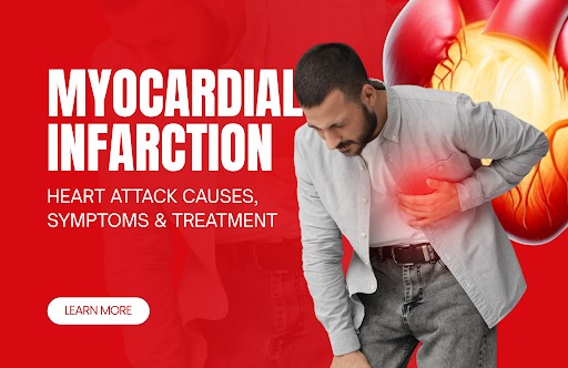
A myocardial infarction occurs when blood supply to a portion of the heart muscle is suddenly cut off. The heart is supplied by the coronary arteries — a network of vessels that sit on the heart’s surface and deliver the oxygen-rich blood the muscle needs to keep beating. When one of those arteries becomes blocked, the heart muscle it supplies begins to die within minutes.

The blockage is almost always caused by coronary artery disease. Over years, fatty deposits called plaque build up inside the coronary artery walls — a process known as atherosclerosis. When a plaque ruptures suddenly, the body’s clotting system responds, forming a blood clot at the rupture site. That clot can block the artery entirely, triggering a heart attack.

Time is muscle. Every minute the artery remains blocked, more cardiac muscle dies — and that damage is permanent. This is why the speed of recognition, emergency response, and intervention determines the long-term outcome as much as any specific treatment decision.

Recognising the Symptoms of a Heart Attack Causes

Heart attacks do not always look the way they do in films. While the dramatic crushing chest pain that radiates to the left arm is a genuine presentation, it is far from the only one. Recognising the full range of symptoms — and taking them seriously — is the first step toward acting in time.

  1. Chest pain or pressure is the most common symptom. Patients describe it as a heaviness, tightness, squeezing, or burning sensation in the centre of the chest. The pain may radiate to the left arm, shoulder, neck, jaw, or back. It typically lasts more than a few minutes, or it may ease and return. A cold sweat, nausea, or dizziness frequently accompanies it.
  2. Shortness of breath may occur alongside chest discomfort or, in some cases, as the dominant symptom. When the heart cannot pump efficiently due to a blocked artery, the lungs begin to back up — and the patient feels breathless, even at rest.
  3. Fatigue and dizziness are symptoms that are easily dismissed, particularly in younger patients and women. A sudden and unexplained exhaustion, or lightheadedness without obvious cause, can be an early signal that the heart is under serious stress.
  4. Nausea and vomiting appear in a meaningful proportion of heart attack patients, particularly those with inferior wall infarctions. These symptoms add to the confusion at presentation, sometimes leading patients — and even non-specialist doctors — to attribute the episode to a gastrointestinal cause.

Any of these symptoms, appearing suddenly and without a clear explanation, should prompt an immediate call to emergency services. They should never be waited out at home.

The Silent Heart Attack: When There Are No Warning Signs

A silent myocardial infarction is one that occurs with no obvious or recognisable symptoms. The patient does not experience chest pain, breathlessness, or any of the classical warning signs. The heart attack happens — muscle is lost — and the patient remains unaware until an ECG or cardiac investigation reveals old damage weeks, months, or years later.

Silent heart attacks are more common in patients with diabetes, in older adults, and in women. They are not minor events. They carry the same risk of future heart failure, arrhythmia, and death as symptomatic infarctions — and because they go unrecognised, they are often untreated. Regular cardiac check-ups are therefore not optional for high-risk individuals; they are essential.

Who Is at Risk and Why

Myocardial infarction is rarely a random event. In the overwhelming majority of cases, a heart attack is the endpoint of a disease process that began years or decades earlier — one that is driven by identifiable and modifiable risk factors.

  1. High blood pressure damages the lining of coronary arteries over time, accelerating the formation of plaque and increasing the risk of plaque rupture. It is one of the most prevalent and under-treated risk factors for heart attack in India.
  2. High cholesterol, particularly elevated LDL cholesterol, directly feeds the atherosclerotic process. Fatty deposits cannot build up in arterial walls without the raw material that circulating LDL provides.
  3. Smoking damages blood vessels, increases clotting tendency, reduces oxygen-carrying capacity of the blood, and accelerates coronary artery disease at every stage. There is no safe level of tobacco use when it comes to cardiovascular risk.
  4. Diabetes and insulin resistance — conditions with extraordinarily high prevalence in India — damage coronary arteries through multiple mechanisms, and diabetic patients with heart attacks tend to present atypically and fare worse without aggressive management.
  5. Sedentary behaviour reduces cardiovascular fitness, worsens cholesterol profiles, and contributes to weight gain, hypertension, and insulin resistance — creating a cluster of risk factors that compounds over time.

Managing these risk factors — ideally with the guidance of a heart Hospital in Hyderabad — is the most effective form of heart attack prevention available.

Types of Myocardial Infarction

Not all heart attacks are the same, and the distinction between types matters for treatment decisions.

STEMI (ST-elevation myocardial infarction) is the most severe form. The coronary artery is completely blocked, blood flow to the affected muscle has ceased entirely, and the ECG shows characteristic ST elevation. STEMI is a cardiac emergency requiring the fastest possible intervention — ideally primary coronary angioplasty with stent implantation within 90 minutes of first medical contact.

NSTEMI (Non-ST-Elevation Myocardial Infarction) involves partial rather than complete arterial blockage. Blood flow is reduced but not fully stopped. The ECG does not show ST elevation, but blood tests reveal elevated troponin — confirming that cardiac muscle is being damaged. NSTEMI requires urgent assessment and typically leads to coronary angiography and intervention within 24 to 72 hours.

How Heart Attacks Are Diagnosed

The diagnostic process combines clinical assessment, electrical recordings, and biochemical testing.

An electrocardiogram (ECG) records the heart’s electrical activity and remains the most important immediate diagnostic tool. In STEMI, characteristic changes allow diagnosis within minutes. In NSTEMI, the ECG may show subtler abnormalities, or may be normal, requiring blood tests to confirm damage.

Troponin blood tests measure the level of troponin — a protein released by damaged cardiac muscle cells — in the bloodstream. Elevated troponin confirms that heart muscle injury has occurred. Serial troponin measurements, taken a few hours apart, improve diagnostic accuracy and help quantify the extent of damage.

Imaging studies, including echocardiography and cardiac CT or MRI, provide additional information about heart function, the location of damage, and the presence of complications. Coronary angiography — an X-ray procedure in which dye is injected directly into the coronary arteries — defines the anatomy of the blockage and guides the interventional plan.

Treatment: Restoring Blood Flow Before More Muscle Is Lost

The primary goal in treating a myocardial infarction is reperfusion — reopening the blocked artery and restoring blood flow to the dying muscle as quickly as possible.

Percutaneous Coronary Intervention (PCI) is the preferred treatment for both STEMI and NSTEMI wherever it can be delivered within the required time window. Using a catheter inserted through the wrist or groin, the interventional cardiologist navigates to the blocked coronary artery under X-ray guidance, inflates a tiny balloon to compress the obstructing plaque, and deploys a PTCA stent — a small mesh cylinder — to hold the artery open permanently. Drug-eluting stents release medication over months to prevent re-narrowing at the treated site. For complex lesions and high-risk patients, chip angioplasty techniques and intravascular imaging are used to optimise stent placement with precision.

Medications form the backbone of both acute and long-term management. In the acute phase, aspirin and anticoagulants prevent further clot formation. Beta-blockers reduce heart workload. ACE inhibitors protect the heart muscle and prevent progressive dysfunction. Statins lower cholesterol aggressively and stabilise remaining plaques against future rupture.

Coronary Artery Bypass Grafting (CABG) is recommended when the anatomy of disease is too complex or extensive for stenting to be the best option — particularly in patients with multi-vessel disease, left main stem disease, or significant diabetes. In CABG, a healthy blood vessel taken from the chest wall or leg is used to reroute blood around the blocked segment, restoring flow to the affected territory.

Prevention: The Work That Happens Before the Heart Attack

The best heart attack is the one that never happens. Prevention is not passive — it requires active management of the risk factors outlined above, regular monitoring, and honest engagement with a cardiologist who will not simply reassure a patient because their resting ECG looks normal.

A healthy, plant-forward diet low in processed foods, salt, and saturated fat supports blood pressure, cholesterol, and weight management simultaneously. At least 150 minutes of moderate aerobic exercise per week is the minimum evidence-based target. Blood pressure, cholesterol, and blood glucose should be checked regularly — and when they are abnormal, treated with appropriate medication, not just lifestyle advice alone.

Stress management is not optional. Chronic psychological stress activates inflammatory pathways, raises blood pressure, and increases heart rate — all of which accelerate coronary artery disease and raise the short-term risk of plaque rupture. Structured techniques — regular physical activity, sleep hygiene, and social connection — are all evidence-based stress reduction strategies.

Conclusion

A myocardial infarction is one of the most time-sensitive medical emergencies in existence. The cardiac muscle that is lost in the first 90 minutes of a heart attack will never regenerate. But the majority of heart attacks are preventable through systematic risk factor management — and when they do occur, outcomes today are dramatically better than they were even a decade ago, thanks to advances in coronary angioplasty, coronary stent technology, and evidence-based cardiac care.

Knowing the symptoms, acting fast, and working with an experienced heart specialist in Hyderabad who manages both prevention and intervention — that combination offers the best possible chance of a full and lasting recovery.

Also Read:

FAQs

What is a myocardial infarction?
A myocardial infarction is the medical term for a heart attack — an event in which a coronary artery becomes blocked, cutting off blood supply to a section of the heart muscle. The affected muscle begins to die within minutes. Prompt intervention by an experienced cardiac team — using coronary angioplasty or other reperfusion strategies — is essential to limit permanent damage.

What are the warning signs of a heart attack Causes?
The most common warning signs are chest pain or pressure, shortness of breath, pain radiating to the arm, jaw, or back, unexplained fatigue, dizziness, nausea, and cold sweating. Some heart attacks, particularly in diabetic patients, present silently or with minimal symptoms. Any unexplained combination of these symptoms should prompt an immediate call to emergency services.

How is a heart attack treated in India today?
The standard treatment for a STEMI heart attack is primary PCI — emergency coronary angioplasty with stent implantation — performed as rapidly as possible after the onset of symptoms. Leading cardiac centres in India achieve door-to-balloon times that match international benchmarks. NSTEMI is managed with urgent angiography and intervention within 24 to 72 hours. Medications are used alongside procedural treatment and continued long-term to reduce the risk of a second event.

How can a heart attack be prevented?
Controlling blood pressure, cholesterol, and blood sugar — through lifestyle and medication where needed — is the foundation of prevention. Stopping smoking, maintaining a healthy weight, exercising regularly, and managing stress all reduce risk significantly. Regular cardiac check-ups with a qualified heart doctor are essential for anyone with risk factors, a family history of heart disease, or a previous cardiac event.

Abhijit

PTCA-STENT-CORONARY-ANGIOPLASTY-EXPERT.webp

February 24, 2026

PTCA Stent जब किसी व्यक्ति को coronary artery disease (हृदय की धमनियों में रुकावट) के बारे में पता चलता है, तो चिंता होना स्वाभाविक है। धमनियों में ब्लॉकेज होने से हार्ट अटैक का खतरा बढ़ जाता है। ऐसे में एक अनुभवी coronary artery disease doctor या coronary artery disease specialist सही समय पर उपचार करके रक्त प्रवाह को सामान्य कर सकते हैं।

भारत में एक अनुभवी coronary heart disease doctor द्वारा की जाने वाली Coronary Angioplasty, Angioplasty in Heart, और PTCA Stent प्रक्रिया ब्लॉकेज को सुरक्षित तरीके से खोलने में मदद करती है। अगर आप Best Angioplasty Expert in India या Angioplasty Expert in India की तलाश में हैं, तो इस गाइड से आपको पूरी जानकारी मिलेगी।

PTCA Stent क्या होता है?

PTCA Stent एक छोटा जालीदार (mesh) ट्यूब होता है जिसे Angioplasty and Angiogram के दौरान ब्लॉक हुई coronary artery में डाला जाता है। इसका मुख्य उद्देश्य धमनियों को खुला रखना और दिल तक रक्त प्रवाह को सामान्य करना है।

आजकल भारत में Stent Coronary के उन्नत प्रकार जैसे Drug-Eluting Stent का उपयोग किया जाता है, जो दवा छोड़कर दोबारा ब्लॉकेज बनने से रोकता है। कई जटिल मामलों में आधुनिक तकनीक जैसे chip angioplasty का भी उपयोग किया जाता है।

Coronary Angioplasty क्यों की जाती है?

जब धमनियों में चर्बी (Plaque) जमा हो जाती है, तो उसे coronary artery disease कहा जाता है। इससे सीने में दर्द (Angina), सांस फूलना और हार्ट अटैक का खतरा बढ़ सकता है।

Coronary Angioplasty एक minimally invasive प्रक्रिया है जिसमें:

  • पहले Angioplasty and Angiogram द्वारा ब्लॉकेज की पहचान की जाती है
  • फिर एक बैलून कैथेटर के माध्यम से नस को खोला जाता है
  • आवश्यकता होने पर PTCA Stent या Stent Coronary लगाया जाता है
  • इस प्रक्रिया को आम भाषा में Angioplasty in Heart भी कहा जाता है।

प्रक्रिया और रिकवरी

  • सबसे पहले कार्डियक कैथेटराइजेशन किया जाता है।
  • एंजियोग्राम द्वारा ब्लॉकेज की जगह पता की जाती है।
  • बैलून फुलाकर नस को खोला जाता है।
  • जरूरत होने पर PTCA Stent लगाया जाता है।


अधिकांश मरीज 1–2 दिन में अस्पताल से छुट्टी पा लेते हैं। हालांकि, कुछ हफ्तों तक भारी काम से बचना चाहिए। सही दवाइयों और जीवनशैली में बदलाव से coronary artery disease को नियंत्रित रखा जा सकता है।


सही विशेषज्ञ कैसे चुनें?


एक अनुभवी coronary artery disease doctor या coronary artery disease specialists का चयन बहुत महत्वपूर्ण है। ध्यान रखें:

✔ डॉक्टर को Coronary Angioplasty और PTCA Stent में अनुभव हो

✔ अस्पताल मान्यता प्राप्त (Accredited) हो

✔ आधुनिक सुविधाएँ और कैथ लैब उपलब्ध हों

✔ मरीजों के अच्छे रिव्यू हों

यदि आप जटिल ब्लॉकेज के लिए Best Angioplasty Expert in India या अनुभवी Angioplasty Expert in India की तलाश में हैं, तो विशेषज्ञ का अनुभव और अस्पताल की सुविधाएँ सफलता दर को बेहतर बनाती हैं।


You can also read this : Angioplasty and Stent: Understanding the Link

Yashoda Hospitals में अनुभवी चिकित्सा स्टाफ प्रत्येक परामर्श के दौरान उन्हें उच्च गुणवत्ता वाली सहायता और रोगी-केंद्रित सेवाएँ प्रदान करता है। आज ही Dr. C. Raghu, जो हैदराबाद के एक अग्रणी हृदय विशेषज्ञ हैं, से अपनी अपॉइंटमेंट बुक करें और Yashoda Hospitals, Somajiguda में सुरक्षित और मजबूत हृदय स्वास्थ्य की दिशा में पहला कदम उठाएँ।


निष्कर्ष

coronary artery disease एक गंभीर स्थिति है, लेकिन सही समय पर उपचार और अनुभवी coronary heart disease doctor द्वारा की गई coronary angioplasty से मरीज सामान्य जीवन जी सकता है। हमेशा प्रमाणित अस्पताल में अनुभवी coronary artery disease specialists या Best Angioplasty Expert in India से परामर्श लें और अपने हृदय स्वास्थ्य को प्राथमिकता दें।


FAQs

  1. PTCA Stent क्या है?

यह एक जालीदार ट्यूब है जो Coronary Angioplasty के दौरान ब्लॉक धमनियों को खुला रखने के लिए डाला जाता है।

  1. Angioplasty and Angiogram में क्या अंतर है?

Angiogram ब्लॉकेज की जांच के लिए होता है, जबकि Angioplasty in Heart ब्लॉकेज को खोलने के लिए की जाती है।

  1. क्या chip angioplasty सुरक्षित है?

हाँ, यह जटिल और कठोर ब्लॉकेज में उपयोग की जाने वाली उन्नत तकनीक है।

  1. क्या Stent Coronary हमेशा जरूरी होता है?

अधिकांश मामलों में नस को खुला रखने के लिए PTCA Stent लगाया जाता है।

Abhijit

IVUS-Guided-Multi-vessel-CHIP-case-with-Rotashock-3.webp

January 16, 2025

IVUS Guided Multi Vessel CHIP Case With Rotablator

Clinical Presentation

  • A 77 years, Male presented with severe angina

Findings

  • Patient is with old CVA history presented with Anterior non-ST Elevation Myocardial Infraction

2D ECHO Findings

  • Mild Concentric LVH+RWHA of LV + Fair LV systolic function
  • Grade 1 LV Diastolic Dysfunction
  • Trivial MR/TR/No PAH
  • No PE/Clot
  • HR-54 BPM

Plan of Procedure:

PTCA and Stent to LAD, LCX and Left Main Coronary Artery Rotablation with IVUS + IVL with IABP support

Abhijit


Dr raghu logo


Dr raghu logo


Call us now if you are in a medical emergency need, we will reply swiftly and provide you with a medical aid.


For Appointment with Dr Raghu: +91 80085 36699
Dr Raghu’s Patient Coordinator: +91 72860 10203
Dr Raghu’s Office Experience: +91 90001 65962
Dr Raghu’s Social Media Communication Channel: +91 97046 51708

Dr. Raghu | Heart Specialist in Hyderabad
Yashoda Hospitals, Raj Bhavan Road, Somajiguda, 1st floor, Room No. 115, Hyderabad -500082, Telangana

Best Cardiologists in Hyderabad


© 2024, Dr. Raghu. All rights reserved. Design & Developed by AMSDigital.in

[gtranslate]
+91-9542475650