heart-attack.jpg

June 6, 2026 AbhijitHealthcare

Heart doctor in Hyderabad are increasingly seeing a troubling trend among younger adults. You are 34, a non-smoker, not diabetic, and seemingly fit. You hit the gym three times a week and eat reasonably well. And then one Tuesday morning, mid-workout, your chest tightens. By the time the ambulance arrives, you are having a massive cardiac event. This is not a rare story anymore. Heart attacks in young people — in their 30s and early 40s — are rising at a rate that has even the most seasoned Heart Specialist in Hyderabad concerned. 

India’s Young Heart Problem Is Real

India has one of the highest rates of premature heart disease in the world. Studies show that nearly 25% of all heart attack deaths in India occur in people under 40. This is not genetics alone. This is a lifestyle crisis. The Indian urban professional is under enormous pressure — career targets, long commutes, rising EMIs, social comparison on Instagram, and a food culture that has quietly shifted from home-cooked dals and rotis to Zomato orders every other night.

Also read: Best Heart Doctor in Hyderabad, Why Dr C Raghu 

Why a Heart Attack at the Gym Is More Common Now

A heart attack in gym settings among young people is not because exercise is dangerous. It is because people with undetected arterial plaque push their hearts into maximum exertion without ever being screened. When you sprint on a treadmill with a partially blocked artery, the sudden surge in demand can rupture that plaque. The resulting clot blocks blood flow — that is a heart attack.

A famous cardiologist in India, like Dr C. Raghu, sees this pattern regularly — young patients who looked completely healthy on the outside but had arteries that were silently narrowing for years. Consulting a trusted top heart specialist in Hyderabad before symptoms appear is the most effective form of prevention.

What Is a Silent Heart Attack?


A silent heart attack causes minimal or no symptoms. You might feel a bit tired for a few days. Some mild jaw, upper back, or arm discomfort. A passing nausea. And then you go back to work. It can cause permanent damage to your heart muscle without you even knowing it happened — often only showing up when you get an ECG for an unrelated reason.

What Is a Minor Heart Attack?

A minor heart attack — medically called an NSTEMI — is often dismissed as indigestion or stress. But it still injures your heart. Each one increases your risk of a more severe event. Any cardiac event is a medical emergency. Time is muscle.

The Lifestyle Changes That Actually Matter


Stop Treating Weekdays as a Write-Off

Your coronary arteries do not work on a Monday-to-Friday write-off schedule. Chronic inflammation from daily junk food, late nights, and inactivity builds the plaque that eventually ruptures.

Understand Your Family History

If your father had a heart attack at 50 or your uncle was on cardiac medication at 45, your risk is significantly higher. This is not destiny — it is a warning to act earlier. Tell your heart doctor in Hyderabad about your family history at every visit.

Get a Cardiac Screening, Not Just a General Health Check

A standard corporate health check measures your weight, blood pressure, and blood sugar. A proper cardiac screening includes an ECG, echo, lipid profile, and sometimes a treadmill stress test — giving a far more complete picture of your heart’s actual health.

Balloon Mitral Valvotomy: When Valve Disease Enters the Picture

Some young Indians — especially women with a history of rheumatic fever — develop valve problems that mimic heart disease symptoms. Balloon Mitral Valvotomy is a minimally invasive procedure used to treat mitral valve stenosis, which can cause breathlessness and fatigue identical to what many young professionals attribute to being out of shape.

Also read: Atrial Fibrillation treatment in India: Complete Guide 

Conclusion

Your heart does not care that you are 34 and busy. If you are an urban professional living a high-pressure, low-movement life, the time to act is right now — not after a cardiac event.

Dr C. Raghu — Interventional Cardiologist, Yashoda Hospital, Somajiguda, Hyderabad
Recognized widely recognised as a famous cardiologist in India for his expertise in complex coronary interventions and preventive cardiology. He has helped young patients across Hyderabad, Bangalore, and beyond reclaim their heart health before a crisis arrives.


pulmonary-embolism-in-india.webp

June 1, 2026 AbhijitHearty Life

A Clot That Travels to the Lung  and the Urgency It Demands

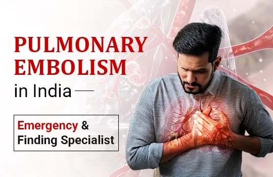
Pulmonary embolism (PE) is a sudden blockage of one or more pulmonary arteries, the blood vessels carrying blood from the heart to the lungs, by a clot that has usually traveled from the deep veins of the legs or pelvis. It is a potentially catastrophic emergency. A massive pulmonary embolism, in which a large clot obstructs the main pulmonary arteries, can produce acute right heart failure, hemodynamic collapse, and cardiac arrest within minutes. Even smaller emboli, if not recognized and treated, can be fatal.

In India, pulmonary embolism is substantially underdiagnosed. Limited awareness of its symptoms among patients and families, incomplete recognition among some clinicians, and the incorrect perception that PE is primarily a Western disease all contribute to diagnostic delays that cost lives. Improving awareness of pulmonary embolism — who is at risk, what it feels like, and what treatment options are available from a qualified pulmonary embolism specialist in India  is therefore genuinely life-saving.

Deep Vein Thrombosis and Pulmonary Embolism: The Same Condition, Different Stages

DVT treatment and pulmonary embolism management are two aspects of managing a single disease: venous thromboembolism (VTE). Deep vein thrombosis (DVT) is the formation of a blood clot in a deep vein, most commonly the deep veins of the calf, thigh, or pelvis. When a piece of that clot breaks off and travels through the venous system to the right heart and into the pulmonary arteries, pulmonary embolism results.

DVT classically presents with unilateral leg swelling, pain, tenderness, and redness, though a substantial proportion of DVT cases are asymptomatic and discovered only when PE occurs. Any asymmetric leg swelling in a patient with risk factors deserves urgent evaluation. Doppler ultrasound of the leg veins, combined with D-dimer blood testing, is the standard diagnostic pathway.

The risk factors for venous thromboembolism in India’s patient population are highly prevalent: prolonged immobility (hospitalization, long-haul flights, bed rest), recent major surgery (particularly hip and knee replacement), active cancer, pregnancy, oral contraceptive or hormone therapy use, obesity, and inherited thrombophilia (blood clotting disorders). Many patients who develop PE have two or more of these factors coexisting.


Read more: DVT to Pulmonary Embolism: The Risks and Prevention 

Recognising Pulmonary Embolism: Symptoms That Demand Immediate Action

Pulmonary embolism symptoms vary with the size of the embolism and the patient’s underlying cardiopulmonary reserve:

Sudden breathlessness is  often the most prominent symptom, appearing abruptly and without obvious cause. Any unexplained acute onset of breathlessness, particularly in a patient with PE risk factors, must be evaluated urgently for pulmonary embolism.

Pleuritic chest pain is  sharp chest pain that worsens with breathing or coughing, resulting from inflammation of the pleura (lung lining) adjacent to the embolic event.

Rapid heart rate (tachycardia) — nearly universal in significant PE, reflecting the cardiovascular stress of reduced pulmonary circulation.

Hemoptysis,  coughing blood-tinged sputum, occurs in pulmonary infarction when a segment of lung tissue loses blood supply.

Dizziness, presyncope, or syncope are  symptoms of hemodynamic compromise, indicating significant right heart strain.

In massive PE, a large central clot causes hemodynamic collapse the presentation is dramatic: severe breathlessness, hypotension, pallor, cyanosis, and potentially cardiac arrest. This constitutes an extreme cardiac emergency requiring immediate transfer to a hospital with pulmonary embolism treatment capabilities and, in appropriate cases, emergency mechanical intervention.

Any combination of sudden unexplained breathlessness, tachycardia, and recent leg swelling should trigger immediate medical evaluation. Delay is dangerous.

Read more: Dr. C. Raghu is one of the best doctors for TAVI/TAVR in Hyderabad  

Diagnosing Pulmonary Embolism

Diagnostic probability is first assessed clinically; validated scoring tools like the Wells PE Score quantify the pre-test probability of PE based on symptoms, signs, and risk factors. In low- to intermediate-probability patients, a D-dimer blood test can safely exclude PE when negative. In high-probability patients, imaging proceeds directly.

CT pulmonary angiography (CTPA) is the gold standard investigation; it demonstrates clots directly in the pulmonary arteries, defines the anatomical extent of embolic burden, and characterizes right heart strain on the same scan. Echocardiography provides rapid bedside assessment of right ventricular function in hemodynamically unstable patients who cannot be safely transported for CTPA.

Treatment: Stratified by Severity

Low-risk PE  normal blood pressure, and no right heart strain  is treated with anticoagulation. Direct oral anticoagulants (DOACs), particularly rivaroxaban and apixaban, are now first-line treatment, enabling outpatient or brief inpatient management for most low-risk cases.

Submassive PE with  normal blood pressure but echocardiographic or CT evidence of right ventricular dysfunction requires close monitoring and anticoagulation, with selective use of catheter-directed therapy for patients who deteriorate or are at high risk of clinical decompensation.

Massive PE  hemodynamic compromise constitutes an emergency requiring immediate restoration of pulmonary blood flow. Systemic thrombolysis (intravenous clot-dissolving drugs) is the primary intervention when not contraindicated. When systemic thrombolysis is contraindicated by bleeding risk or has failed to produce clinical improvement, catheter-based mechanical thrombectomy physical removal or fragmentation of a clot from the pulmonary arteries—is the advanced intervention that can be life-saving.


Read more: Hyderabad Cardiologist Reveals 5 Silent Heart Warning Signs  

Mechanical Thrombectomy: Advanced PE Care Available in India

Mechanical thrombectomy for pulmonary embolism involves navigating a catheter through the femoral vein, through the right heart, and into the pulmonary arteries, where dedicated devices aspirate or fragment the clot and restore blood flow without the systemic bleeding risk of intravenous thrombolytics. Large-bore aspiration thrombectomy systems and ultrasound-facilitated catheter-directed thrombolysis are the principal modalities used.

This advanced intervention requires specific training, a specialized catheterization laboratory team, and the rapid mobilization of a multidisciplinary PE Response Team (PERT). Dr. Raghu’s practice in Hyderabad maintains this PE treatment capability, offering patients with high-risk PE access to the most advanced catheter-based interventional options available.

For PE evaluation, DVT treatment, or urgent vascular consultation, contact Dr. Raghu’s team. For active emergencies, contact emergency services immediately.
Call Us: +91 95424 75650
Write to us: secretary@drraghu.com


top-heart-doctor-in-hyderabad-1200x778.jpg

May 26, 2026 AbhijitHearty Life

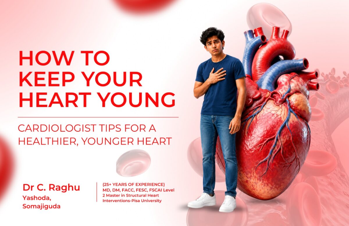
Cardiologist tips check your phone, skip breakfast, and rush through traffic to reach the office. After sitting for long hours, stress, unhealthy food, and lack of exercise become part of daily life. According to a leading cardiologist in Hyderabad, this modern corporate lifestyle is silently increasing the risk of heart disease among working professionals in cities like Mumbai, Bangalore, Pune, and Hyderabad. Millions are living this routine every day — and their hearts are paying the price silently. 

The problem is not that you don’t care about your heart. The problem is that you think you still have time.

Why Your Heart Ages Faster Than You Think

Your heart is a muscle. Like every other muscle, it thrives on movement. It needs good nutrition and rest. But when you spend 9 to 10 hours sitting every day, eat high-salt, high-fat takeout food, and carry the weight of professional deadlines, your heart goes through a kind of invisible wear and tear. Which the cardiologists call cardiovascular ageing, and it begins much earlier than most people expect.

By the time you are in your late 30s or early 40s, years of sedentary habits, stress, and poor diet have already left marks on your arteries. The good news is you can slow this process. A skilled heart doctor in Hyderabad will tell you the same thing: the heart responds well to the right care, at any age.

The Biggest Threats to Your Heart Right Now

  • Chronic sitting: Sitting for more than 6 hours a day increases cardiovascular disease risk by up to 64%, even if you work out on weekends.
  • High-stress environment: Prolonged work stress raises cortisol, which raises blood pressure and inflammation, both major risk factors for heart disease.
  • Poor food choices: Office cafeteria meals and food delivery apps have made it easy to eat processed, calorie-dense, nutrient-poor food every single day.
  • Disrupted sleep: Late nights on OTT platforms mean your heart never gets the deep recovery time it needs.
  • Ignoring early signs: Occasional chest tightness, breathlessness when climbing stairs, or unexplained fatigue are signals many people explain away.
     

Also read:  Dr. C. Raghu is one of the best doctors for TAVI/TAVR in Hyderabad 

What Cardiologist tips Recommend

1. Move More — Even at Your Desk

You do not need to become an athlete. A heart specialist in Hyderabad will tell you that even 30 minutes of brisk walking, five days a week, has measurable benefits on cholesterol, blood pressure, and heart function. If you work from home, set a timer to stand up and walk for 5 minutes every hour.

2. Watch What You Eat on Weekdays

Most of us eat reasonably well on weekends but switch to chaos during our weekdays. Our heart does not get a holiday. Just swap one meal a day to something heart-friendly — maybe a dal and sabzi thali, fresh fruit, or a handful of mixed nuts. We need to reduce sodium by cutting packaged snacks.

3. Know Your Numbers

When did you last check your blood pressure, blood sugar, and cholesterol? These three numbers tell a Heart Specialist in Hyderabad more about your cardiac risk than how you feel right now. You should have a routine lipid profile and ECG checks. It can spot trouble before it becomes a crisis.

4. Manage Stress Like a Skill

Many times, our stress becomes unavoidable due to our work culture. But how you manage it well determines how much damage it does. Even 10 minutes of deep breathing, a short walk after lunch, or a weekend away from screens significantly lowers cortisol. Sleep is not a luxury; it is cardiac medicine.

5. Do Not Wait for Symptoms

Heart disease is notoriously sneaky. Many people who suffer a heart attack have no classic warning signs. If you are over 35, overweight, or have a family history of heart disease, see a heart expert in Hyderabad proactively. A preventive consultation is far less expensive than an emergency.

Also read: Daily Habit That May Double Heart Attack Risk, Warns Cardiologist 

Red Flags You Should Never Ignore

You need to see the best cardiologist in Hyderabad immediately if you experience:

  • Chest pain or tightness, even brief or mild
  • Shortness of breath while doing simple tasks
  • Palpitations or a racing heart without an obvious reason
  • Dizziness or sudden fatigue
  • Swollen ankles or feet

Conclusion

Your heart has been working for you every second of your life. It is time to work for it. Whether you are in Hyderabad, Bangalore, Mumbai, or Chennai, schedule that overdue check-up with a cardiologist. Do not wait for a warning. Make an appointment with Dr Raghu

Dr C. Raghu — Interventional Cardiologist, Yashoda Hospital, Somajiguda, Hyderabad
One of India’s most respected interventional cardiologists with decades of experience in complex cardiac interventions and preventive cardiology. Dr C Raghu has helped thousands of patients across India take control of their heart health — before and after a cardiac event.
Call Us :+91 95424 75650 | Email :secretary@drraghu.com


best-heart-doctor-in-hyderabad.webp

May 19, 2026 AbhijitHearty Life

Best heart doctor Comprehensive Cardiac Care — Why It Matters for the Patient

Cardiac conditions do not present in isolation. A patient with best heart doctor in Hyderabad may simultaneously have atrial fibrillation. A patient with severe aortic stenosis also needs coronary assessment before any valve intervention. A patient with pulmonary embolism may have an underlying thrombophilia that changes the long-term anticoagulation management entirely. A patient with peripheral artery disease almost certainly has coronary disease as well.

The best long-term cardiac outcomes come not from isolated specialist interventions delivered in silos but from integrated, comprehensive cardiac care by a single expert who sees the whole picture, manages multiple conditions within a unified clinical framework, and takes long-term responsibility for the patient’s cardiac health.

This is the model of care that Dr. Raghu, a leading top heart specialist in Hyderabad, delivers every day. His clinical expertise is broad, spanning interventional cardiology, structural heart disease, rhythm management, vascular medicine, and preventive cardiology, and his approach to every patient is integrative rather than procedure-focused.

The Clinical Breadth That Sets Dr. Raghu’s Practice Apart

When patients consult Dr. Raghu’s practice in Hyderabad, they access a cardiologist whose comprehensive cardiac services span multiple subspecialties.

Coronary Artery Disease and Angioplasty: From initial risk stratification and non-invasive investigation through coronary angiography and complex coronary angioplasty (PCI), including intravascular imaging-guided procedures, physiological lesion assessment, and chip angioplasty for calcified lesions. Emergency primary PCI for acute myocardial infarction is available around the clock.

TAVR and Structural Heart Disease: Comprehensive evaluation of aortic stenosis and other valvular conditions, with Heart Team-based decision-making for TAVR candidacy, pre-procedural cardiac CT planning, and transcatheter aortic valve implantation for eligible patients. Patients from across India and internationally consult the program for TAVR evaluation and second opinions.

Atrial Fibrillation and Arrhythmia Management: Systematic AFib evaluation including echocardiography, Holter monitoring, stroke risk stratification, evidence-based anticoagulation, and individualized heart rate and rhythm control strategy. Collaborative referral to cardiac electrophysiology for catheter ablation when indicated.

Pulmonary Embolism and DVT: Acute PE diagnosis and risk stratification, anticoagulation management, and advanced catheter-based intervention, including mechanical thrombectomy for high-risk PE. DVT evaluation and long-term VTE management.

Peripheral Artery Disease and Fistuloplasty: Peripheral arterial assessment and endovascular intervention for claudication and critical limb ischemia. Fistuloplasty and fistula rescue services for hemodialysis patients with access dysfunction.

Preventive Cardiology: Cardiovascular risk assessment, lipid management, blood pressure optimization, and structured prevention programs for patients with high-risk profiles or strong family histories.

Also read : Dr. C. Raghu is one of the best doctors for TAVI/TAVR in Hyderabad

What the First Consultation at Dr. Raghu’s Practice Involves

New patients at Dr. Raghu’s practice receive a consultation structured around building a complete understanding of their cardiac history, symptoms, risk factors, and prior investigations before any treatment plan is discussed. A detailed history covers the presenting symptom in full clinical context, the patient’s cardiovascular risk profile, all current medications, and prior cardiac events or investigations. A focused cardiac examination follows, with findings explained clearly to the patient.

From this foundation, investigation planning is tailored to the specific clinical question—not driven by protocol. A patient with exertional chest pain and multiple risk factors needs a different investigation pathway from a patient referred for valvular assessment or rhythm evaluation. The best cardiologist in India does not apply a one-size-fits-all investigation template to every patient; they think about the clinical question and design the most efficient, informative diagnostic pathway.

Results are explained clearly in a follow-up consultation or communication with the clinical context, the implications, and the treatment options all presented in terms the patient and family can act on.

The Preventive Dimension: Getting Ahead of Heart Disease

A significant part of Dr. Raghu’s practice serves patients who do not yet have symptomatic cardiac disease but whose risk factor profile places them at significant future cardiovascular risk. Patients with strong family histories of early heart disease, those with multiple poorly controlled risk factors, and those who have already had a cardiac event and want to minimize the risk of another all benefit from the systematic, evidence-based approach to cardiovascular risk reduction that preventive cardiology provides.

In the Indian population  with its high prevalence of diabetes, hypertension, and genetic susceptibility to coronary disease  preventive cardiology interventions, applied rigorously and early, are among the highest-value activities any heart specialist doctor in Hyderabad can offer.

Long-Term Relationship: The Real Measure of Cardiac Care Quality

The true test of a cardiac practice is not the quality of a single procedure. It is the quality of the long-term relationship  the ongoing availability for questions, the proactive monitoring of disease progression, the timely adjustment of treatment as new evidence emerges or the patient’s circumstances change, and the accessibility of the physician when new symptoms arise.

Dr. Raghu’s practice maintains structured follow-up for all patients with significant cardiac conditions, provides responsive communication for questions between scheduled visits, and takes a proactive approach to reviewing management plans as guidelines evolve. For patients who have moved cities or live overseas, remote consultations enable the continuation of this relationship beyond geography.

New patients, second opinions, and complex referrals are all welcomed at Dr.C Raghu cardiac practice. Contact the team through

Call Us +91 95424 75650 | write to us : secretary@drraghu.com 


Coronary-Artery-Disease-in-India.jpg

May 15, 2026 AbhijitHearty Life

Coronary artery disease (CAD) kills more Indians than any other single condition. It strikes earlier in the Indian population than in Western countries, often a decade sooner,  driven by a combination of genetic susceptibility to dyslipidemia and insulin resistance, high rates of diabetes, rising rates of obesity and physical inactivity, tobacco use, and a diet increasingly influenced by processed food.

Yet the treatment available today — when coronary artery disease is detected early enough and managed by a skilled coronary artery disease specialist — is remarkably effective. Coronary angioplasty in heart arteries, supported by modern drug-eluting stents and evidence-based medication, gives patients with even severe coronary disease a very real path back to normal life.

The barrier is not treatment availability. It is awareness: patients who do not know the symptoms, who dismiss warning signs, or who do not understand what a coronary angioplasty and angiogram involve delay seeking help in ways that cost them heart muscle, cardiac function, and, sometimes, their lives.

This guide walks every reader through the complete pathway from recognizing symptoms through the diagnostic angiogram to PTCA stent placement and recovery—because understanding this pathway is genuinely life-saving.

Also read: Understanding PTCA Stent & Coronary Angioplasty expert

What Happens in Coronary Artery Disease

coronary artery disease

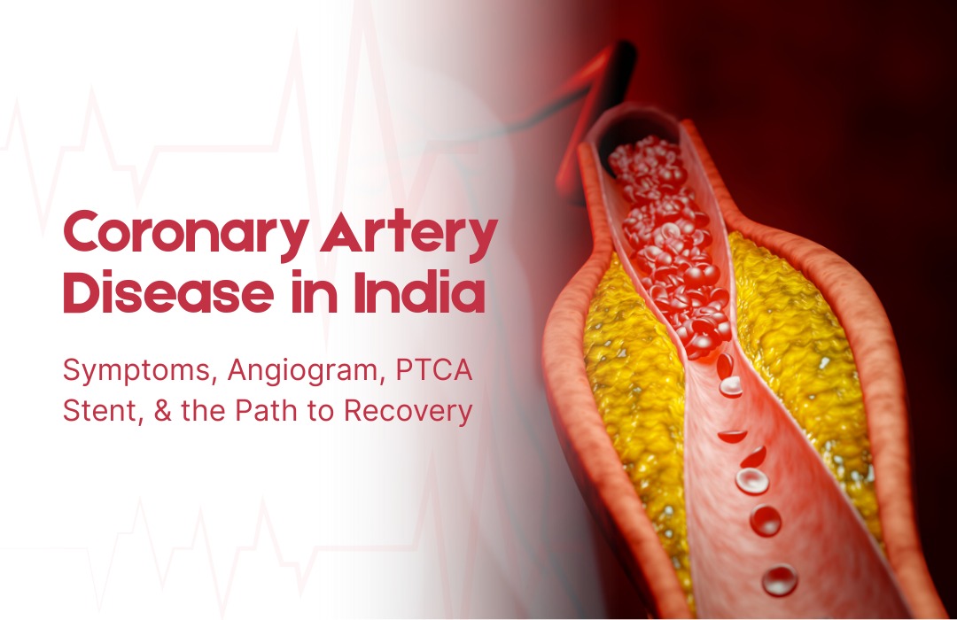
Coronary arteries are the three main blood vessels that supply oxygenated blood to the heart muscle itself. Coronary artery disease is the progressive accumulation of atherosclerotic plaque deposits of cholesterol, inflammatory cells, calcium, and connective tissue within the walls of these arteries. As plaques grow, they narrow the artery’s lumen, reducing the volume of blood the heart muscle receives.

In early disease, there are no symptoms. As narrowings become significant, the heart muscle receives adequate blood at rest but insufficient blood during exertion, producing the symptom of stable angina. When a plaque ruptures suddenly, as can happen without warning at any level of physical exertion, it triggers the formation of an acute blood clot that can completely block the artery, causing a heart attack 

Heart attacks cause permanent heart muscle damage in proportion to the time the artery remains blocked. Opening the blocked artery within 90 minutes of symptom onset with emergency coronary angioplasty (primary PCI) salvages the most muscle and produces the best survival outcomes. Every minute matters.

Recognising the Symptoms: What to Know, and What to Take Seriously

Stable angina is exertional chest discomfort—a tightness, pressure, heaviness, or aching in the center of the chest that appears predictably during physical activity (walking uphill, climbing stairs, carrying loads) and relieves with rest within a few minutes. It may radiate to the left arm, jaw, or back.

In the Indian population, and particularly in patients with diabetes, coronary artery disease sometimes presents atypically without classic chest pain,but with unexplained breathlessness on exertion, unusual fatigue, or a feeling of heaviness or indigestion. These symptoms in any patient with cardiovascular risk factors deserve a prompt cardiac evaluation, not reassurance.

Acute coronary syndromes,  unstable angina and heart attack, present with chest discomfort that is more severe and longer-lasting (more than 20 minutes) and may occur at rest. Associated symptoms, including sweating, nausea, breathlessness, or syncope, increase the likelihood of an acute event. These symptoms require immediate emergency care calling an ambulance and going directly to the nearest hospital capable of emergency coronary angioplasty in heart arteries.

The Diagnostic Pathway: From Symptom to Angiogram

The investigation of stable chest pain begins with clinical history, examination, a resting ECG, and blood tests. An echocardiogram assesses left ventricular function and wall motion. Stress testing exercises, ECG, stress echocardiogram, or nuclear perfusion imaging demonstrates whether coronary disease is causing ischemia during physical stress.

When an investigation suggests significant coronary artery disease, or when a patient presents with an acute coronary syndrome, coronary angiography is performed. This angioplasty and angiogram procedure takes 20 to 40 minutes under local anesthesia and sedation, with a catheter inserted through the radial artery in the wrist to deliver contrast dye into the coronary arteries while X-ray images map the distribution and severity of narrowings.

CT coronary angiography, a noninvasive alternative using CT scanning, is increasingly used as an initial anatomical investigation in stable patients with intermediate pretest probability of significant coronary disease, providing detailed arterial mapping without catheter insertion.

Also read: 5 Signs You or Your Elderly Parent May Need a Heart Valve Replacement  

PTCA Stent: What the Procedure Involves Step by Step

When the angiogram reveals a narrowing that warrants treatment, angioplasty with PTCA stent deployment proceeds as follows:

A guidewire, a very thin, flexible wire, is advanced through the catheter and across the coronary narrowing. The wire serves as a rail for all subsequent equipment. A balloon catheter is advanced over the wire to the lesion site and inflated to compress the plaque, widening the arterial lumen. A drug-eluting stent coronary device, a metal mesh cylinder mounted on a second balloon, is then advanced to the same position and expanded, embedding itself in the artery wall and remaining in place permanently as a scaffold.

After stent deployment, the operator assesses the result using post-deployment balloon inflation to optimize expansion and intravascular imaging (IVUS or OCT) in many cases to confirm optimal stent apposition. Haemodynamic measurements confirm normal blood flow before all equipment is removed.

For heavily calcified lesions that require special preparation before stenting, chip angioplasty techniques, including intravascular lithotripsy or rotational atherectomy, are deployed to modify the calcium and allow adequate stent expansion.
The entire procedure for a straightforward single-vessel case typically takes 45 to 90 minutes.

Recovery: Life After a Coronary Stent

Recovery from coronary angioplasty is rapid compared to surgical alternatives. Most patients return to light daily activities within one to two weeks and to full activity within two to four weeks.

The most critical post-stent requirement is medication adherence. Dual antiplatelet therapy, aspirin combined with a second agent (clopidogrel, prasugrel, or ticagrelor), is prescribed for a defined period to prevent stent thrombosis. The duration depends on stent type and clinical risk profile. Stopping this medication early without medical guidance carries a risk of acute stent occlusion and must be avoided.

Long-term coronary artery disease management, statins, blood pressure treatment, diabetes optimization, and lifestyle modification continue indefinitely regardless of whether a stent has been placed. A stent treats the obstruction; it does not cure the underlying disease. Regular follow-up with the treating coronary artery disease doctor is essential for monitoring disease progression and adjusting treatment as needed.

Dr. C Raghu: Comprehensive Coronary Artery Disease Care in Hyderabad

Dr. C. Raghu coronary artery disease program in Hyderabad encompasses the full clinical pathway from initial risk assessment and non-invasive investigation through coronary angiography and complex coronary angioplasty to long-term secondary prevention management. His practice uses intravascular imaging and physiological assessment tools routinely, reflects current evidence-based guidelines in every decision, and provides clear, patient-centered communication throughout every stage of the diagnostic and treatment process.

For advanced coronary artery disease treatment, coronary angioplasty, and PTCA stent care in Hyderabad, consult Dr. C. Raghu at Yashoda Hospitals, Secunderabad — Call +91 95424 75650 today for expert cardiac care.


Best-Angioplasty-Expert-in-India.webp

May 13, 2026 AbhijitHearty Life

India’s Coronary Disease Crisis — and Why the Quality of the Doctor Matters

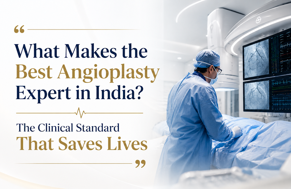
Angioplasty expert in India Coronary artery disease is the leading cause of death in India. The country bears one of the world’s highest burdens of coronary disease, presenting at younger ages than Western populations and frequently at more advanced stages of disease. Every year, hundreds of thousands of Indians undergo coronary angioplasty, the catheter-based procedure to open blocked heart arteries, as a treatment for chest pain, heart attacks, and significant coronary disease discovered on investigation.

The outcome of that angioplasty, whether it relieves symptoms effectively, whether the stent lasts, whether the vessel stays open, or whether complications are managed safely, depends enormously on the quality of the operator. Not all interventional cardiologists are equal. The gap between the best angioplasty experts in India and average operators is meaningful, and that gap is most visible in exactly the cases where it matters most: complex lesions, challenging anatomy, and patients with multiple co-existing conditions.

This article explains what genuinely separates the best angioplasty experts in India from the rest — using plain language, grounded in evidence, because patients who understand these distinctions make better decisions about their own care.

What Coronary Angioplasty Actually Is

Coronary angioplasty, formally called Percutaneous Coronary Intervention (PCI) and historically known as PTCA (Percutaneous Transluminal Coronary Angioplasty), is a catheter-based procedure to open narrowed or blocked coronary arteries that supply blood to the heart muscle.

Under local anesthesia and light sedation, a catheter is inserted through the radial artery in the wrist (the modern preferred access route) or the femoral artery in the groin and navigated under X-ray guidance to the blocked coronary artery. A tiny balloon is inflated at the site of the blockage to compress the obstructing plaque and widen the artery. A PTCA stent  a small metal mesh cylinder  is then deployed at the same site to hold the artery open permanently.

Modern drug-eluting stents, which release medication over several months to prevent re-narrowing at the treated site, have dramatically improved the long-term success rates of coronary angioplasty. Complex cases involving heavily calcified plaques benefit from specialized techniques, including chip angioplasty using intravascular lithotripsy, cutting balloons, or rotational atherectomy, that the best angioplasty experts in Hyderabad are trained and equipped to deploy.

Also read : Meet Cardiologist in India, Dr C. Raghu Yashoda, Somajiguda 

Differentiator 1: Case Volume and Complexity Range

The relationship between operator volume and patient outcomes in interventional cardiology is one of the most extensively documented findings in all of medicine. Operators who perform a high annual volume of coronary angioplasty procedures make fewer technical errors, handle complications more adeptly, make better judgments about when to intervene and when not to, and produce measurably better outcomes across virtually every clinical metric.

This is particularly important in complex cases. Single-vessel, non-calcified, easily accessed coronary lesions in otherwise healthy patients are manageable for most trained interventionalists. Multivessel disease, chronic total occlusion recanalization, left main coronary stenting, severely calcified lesions requiring chip angioplasty techniques, and patients with prior coronary artery bypass surgery  these cases require accumulated experience that only comes with high-volume practice.

When a patient has complex coronary artery disease, the choice of a high-volume complex PCI operator is not a luxury. It is a clinical necessity that affects outcomes.

Differentiator 2: Intravascular Imaging Guidance

Coronary angiography — the standard X-ray imaging of coronary arteries using contrast dye — shows where narrowings are and how severe they appear. But it provides only a two-dimensional silhouette view of a three-dimensional structure. It cannot reliably assess plaque composition, calcium distribution, stent expansion, or stent apposition to the vessel wall.

Intravascular imaging — specifically intravascular ultrasound (IVUS) and optical coherence tomography (OCT) — provides cross-sectional views inside the artery in high resolution, allowing the angioplasty expert to see inside the vessel in three dimensions, choose the correct stent size, guide deployment, verify optimal expansion, and identify and correct problems before the patient leaves the catheterization laboratory.

Multiple large randomized trials and registry studies have confirmed that intravascular imaging-guided PCI is associated with lower rates of stent thrombosis, lower rates of target vessel failure, and better long-term clinical outcomes compared to angiography-guided PCI alone. An angioplasty expert in India who routinely uses intravascular imaging is practicing at a higher standard than one who relies on angiography alone.

Differentiator 3: Physiological Assessment of Lesion Severity

Not every coronary narrowing visible on an angioplasty and angiogram needs to be treated with a stent. Coronary angiography sometimes makes narrowings look more severe or less severe than they physiologically are. Treating a narrowing that does not cause ischemia (inadequate blood supply to heart muscle) with a stent exposes the patient to the risks of the procedure without providing clinical benefit.

Fractional Flow Reserve (FFR) and instantaneous Wave-Free Ratio (iFR) are wire-based physiological measurements that assess whether a coronary narrowing is causing clinically significant ischemia. Multiple trials — DEFER, FAME, FAME 2, and iFR-SWEDEHEART — have established that FFR- and iFR-guided PCI is superior to angiography-guided PCI in reducing unnecessary procedures while improving outcomes in lesions that truly require treatment.

A coronary heart disease doctor who uses physiological assessment to guide stent decisions is practicing evidence-based medicine at a higher standard than one who relies solely on the visual appearance of angiography.

Differentiator 4: Honest Decision-Making About When Not to Stent

Perhaps the most underappreciated quality of the best angioplasty experts in India is their willingness to recommend against angioplasty when the evidence does not support it. Major trials — ISCHEMIA, COURAGE, BARI 2D — have demonstrated that in patients with stable coronary artery disease and good left ventricular function, optimal medical therapy alone can produce outcomes equivalent to PCI for preventing heart attacks and death, while PCI reliably provides better symptom relief.

A coronary artery disease specialist who presents these findings honestly, explains that a stent will relieve angina but may not prolong life in stable disease, and supports the patient in making an informed choice rather than defaulting to the most immediately available intervention is providing genuinely excellent, patient-centered care.

This kind of honest decision-making is a marker of clinical confidence and integrity that patients should look for and value.

Also read: CHIP Angioplasty: Advanced Coronary Artery Disease Treatment Explained 

Emergency Angioplasty: The Time-Critical Dimension

The most time-sensitive application of coronary angioplasty is primary PCI the emergency procedure to open a blocked artery during an acute heart attack (STEMI ST-elevation myocardial infarction). Primary PCI must be performed within 90 minutes of hospital arrival to minimize heart muscle damage and improve survival. Every minute of delay costs irreplaceable cardiac muscle.

An angioplasty expert in Hyderabad who participates in a 24/7 primary PCI program on call around the clock, working in a center with rapid cath lab activation protocols, demonstrates the kind of commitment to acute cardiac care that defines a truly comprehensive coronary program. Dr. Raghu practice maintains this emergency PCI capability, ensuring that patients with acute coronary syndromes receive the most time-critical interventional care when it is needed most.

Dr. Raghu: The Standard of Excellence in Coronary Angioplasty in Hyderabad

Dr. C Raghu brings to his coronary angioplasty practice a combination of high case volume, routine intravascular imaging and physiological lesion assessment, mastery of complex PCI techniques, including chip angioplasty for calcified lesions, and an evidence-based communication approach that helps patients understand exactly when angioplasty adds value to their treatment and when other options deserve equal consideration.

His patients  from simple stable coronary disease to the most complex multivessel and chronic total occlusion cases — receive a consistently high standard of evaluation, procedural care, and long-term follow-up.

Contact Dr.C Raghu’s team cardiologist in Hyderabad through to schedule a coronary artery disease evaluation or angioplasty consultation.

About Dr. C. Raghu

Dr. C. Raghu — MD, DM, FESC, FACC, FSCAI
Interventional Cardiologist | Yashoda Hospital, Raj Bhavan Road, Somajiguda, 1st Floor, Room No. 115, Hyderabad – 500082, Telangana

Specialisation: Coronary, Vascular & Structural Interventions | TAVR | CHIP Angioplasty | Complex Coronary Procedures

OPD: Monday to Saturday, 8 AM – 8 PM  |  Emergency: 24/7  |  +91 95424 75650  |


Dr raghu logo


Dr raghu logo


Call us now if you are in a medical emergency need, we will reply swiftly and provide you with a medical aid.


For Appointment with Dr Raghu: +91 80085 36699
Dr Raghu’s Patient Coordinator: +91 72860 10203
Dr Raghu’s Office Experience: +91 90001 65962
Dr Raghu’s Social Media Communication Channel: +91 97046 51708

Dr. Raghu | Heart Specialist in Hyderabad
Yashoda Hospitals, Raj Bhavan Road, Somajiguda, 1st floor, Room No. 115, Hyderabad -500082, Telangana

Best Cardiologists in Hyderabad


© 2024, Dr. Raghu. All rights reserved. Design & Developed by AMSDigital.in

[gtranslate]
+91-9542475650