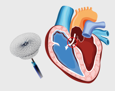
cci-device_closure_for_pda.jpg

January 16, 2021 Dr. RaghuHearty Life

Patent Ductus Arteriosus (PDA) is a congenital heart condition in which the opening between the pulmonary artery and the aorta, persists after its normal closure time. The ductus arteriosus is a small connection between the two major blood vessels in the fetal heart, and it naturally closes shortly after birth. When it fails to close, it’s called patent ductus arteriosu

A PDA may allow the oxygenated blood to mix with the deoxygenated blood, compromising the heart and lung function.

What are the treatment options for PDA?

  • The treatment options depend on the age of the person.
  • In a premature baby, the PDA may close with time, as the baby grows. Some babies may need medications such as indomethacin, to facilitate closure. Surgery may be needed if the duct fails to close.
  • In full-term babies, a small defect can be monitored, as it may close with time. Large defects may need to be closed surgically.
  • Surgical closures can be achieved through open-heart surgery and percutaneous catheterization.

What is Device Closure for Patent Ductus Arteriosus (PDA)?

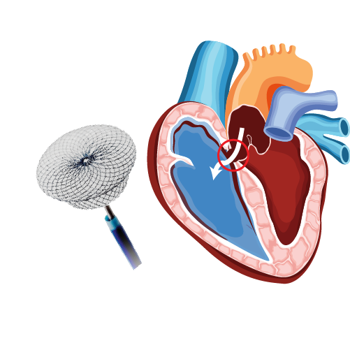
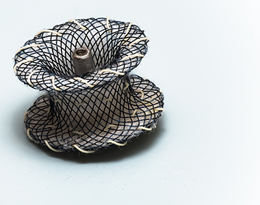
The larger PDA results in increased workload of heart, and also carries a risk of bacterial infection. PDA can be closed by inserting a device through the blood vessel in groin, a non-surgical method called percutaneous transcatheter approach.

Who is eligible for this treatment?

This method is considered only if the child is

  • Older than 6 months
  • At least 22 pounds
  • With defects that are not too large

Considerations in adults include:

  • The device closure is considered as long as the elevated pressure in the lungs is not irreversibly elevated
  • If the lung pressure is already very high, it is carefully measured along with lung resistance to determine the safety the procedure.

What are the risks associated with the procedure?

PDA closure is a low risk procedure, but the common risks include:

  • Rupture of the blood vessel or the heart wall
  • Complications during positioning the device
  • Leakage through the closure device

What happens during the procedure?

  • In case of adults, a sedative might be given to relax them, and a local anesthetic is given to numb the site of catheter introduction.
  • Children are administered general anesthesia during the procedure.
  • A catheter is inserted into the blood vessel in the groin. It is then moved up to the heart into the PDA.
  • The pressure, oxygen saturation and the size of opening in the heart is measured.
  • A closure device is threaded through the catheter and placed onto the PDA.
  • Once the device is in place, it is pushed out of the catheter to implant it over the opening.

What care should be taken post-procedure?

  • After implantation, the catheter is removed, and the incision is closed.
  • The procedure usually takes between 1 and 3 hours.
  • You might need to have an X-ray after the procedure to ensure the implant is at the right position.
  • An echocardiogram may be recommended after six months to ensure that the PDA is properly closed.
  • Antibiotics might be prescribed to prevent endocarditis.
  • The child may need to come for regular follow-ups to ensure that the device closure is effective.

disease-condtions-device_closure_for_asd.jpg

January 16, 2021 Dr. RaghuHearty Life


An atrial septum is a muscular wall that separates the upper chambers of the heart called atria. An ASD is a common congenital heart disease where the septum is not formed properly producing a left-to-right shunt, which leads to mixing of oxygenated and deoxygenated blood. This causes pulmonary hypertension and right heart enlargement.

What are the treatment options for ASD?

Small atrial defects do not need any treatment and close on its own. Even in adulthood small ASDs may remain asymptomatic. Some large defects that persists in adulthood may become symptomatic and need closure. The ASDs can be closed by:

  • Percutaneous closure using a device
  • Closure through open heart surgery

Percutaneous device closure is the preferred treatment for certain defects type.

What is Percutaneous Closure of Atrial Septal Defect (ASD)?

A noninvasive procedure known as percutaneous transcatheter approach is considered depending on the size and severity of the defect. Moderate to large-sized ASD along with pulmonary hypertension requires to be closed. The procedure is performed by inserting a special closure device either folded or attached to a catheter into the vein of the leg and is advanced to the heart through the defect, which closes the hole by a special mechanism.

Are there risks associated with the procedure?

The success rate of the procedure is about 95%. But the risks involved, and their estimated incidence of occurrence include:

  • Device dislodgement leading to emergency heart surgery: 1%
  • Device erosion (device eroding through the heart walls): 0.3%
  • Death: less than 1% usually due to perforation of the heart chamber
  • Dislodgement of clot or air bubbles to the brain leading to other organs: less than 1%
  • Arrhythmia: 1 to 2%
  • Allergic dye reaction
  • Anaesthetic reaction
  • Injury to the artery/vein/nerves in the groin
  • Perforation of the oesophagus
  • Infection
  • Allergic reaction to the nickel component of the device

Patients with small ASD may not develop any complications, but large-sized defects may lead to serious complications which demands surgery and prolonged hospitalization.

What is the pre-procedure work-up?

Pre-operative tests to assess the general health of the patient include:

  • Chest x-ray
  • Electrocardiogram
  • Blood tests
  • Kidney function

A detailed diagnosis of the defect should be performed which includes transthoracic and transoesophageal echocardiogram used to assess the size, location and the suitability of the procedure.

How should I prepare for the procedure?

  • Patients wearing dentures, glasses or a hearing assist device can plan to wear them during the procedure.
  • Patient will be instructed about dietary restriction to be followed before the procedure.

Are there any specific instructions about medications?

  • The healthcare provider may ask you to stop certain medications, such as warfarin or other blood thinners.
  • If diabetic, consult the physician about how the medication needs to be adjusted.
  • Provide information about specific allergies regarding iodine, shellfish, X-ray dye, latex or rubber products etc.

What happens during the procedure?

  • The patient might be asked to have a shower before the procedure.
  • The patient is asked to wear a hospital gown and lie on an X-ray table where an X-ray camera will move over the chest during the procedure.
  • Arrangements for intravenous administration of medications or fluids during the procedure will be made.
  • The site where the catheter will be inserted is cleaned and sterile drapes were used to cover the site to prevent the infection.
  • Electrodes will be placed on the chest and are attached to an electrocardiograph monitor (ECG).
  • A sedative might be given to relax, and a local anesthetic is given to numb the site of catheter introduction.
  • A plastic sheath will be inserted in the groin, through which a catheter is inserted and threaded to the heart.
  • The physician may also inject a dye which may make you feel hot or flushed for several seconds. Inform the doctor if there is an allergic reaction like itching or tightness in throat, nausea and chest discomfort.
  • The X-ray cameras are used to obtain the measurements of pressures and oxygen content in the chambers.
  • The appropriate size and the location of the closure might be visualized using a small catheter connected with an ultrasound transducer.
  • A special catheter is used to advance the device into the heart and through the defect.
  • The device is slowly pushed out of the catheter allowing each side of the device to open and close each side of the hole in the septum.
  • The proper position of the device is ensured and is released from the catheter.
  • It may take 1-2 hours for the procedure, but preparations must be made to spend about 5-9 hours in the hospital.
  • The patient should be accompanied by someone who can drive him home, as the patient will not be allowed to drive on the same day.

What care should be taken after the procedure?

  • The catheter and the imaging probe are removed after the completion of the procedure.
  • Pressure on the incision site or occasionally a small suture is used to close the vein.
  • Bed rest is advised for several hours to prevent bleeding, but call the doctor if you notice any bleeding.
  • You might be advised to drink plenty of water to wash out the contrast material from the body.
  • Your heart rate and rhythm are monitored; you may be asked to stay overnight in the hospital.
  • Medications, such as aspirin are prescribed to prevent blood clots.
  • Strenuous activity and heavy lifting should be avoided for at least six months.
  • Antibiotic prophylaxis is required for at least six months or lifelong to prevent endocarditis, as per doctor’s advice.


  • 24 hours emergency services
24 hours emergency services
  • Clinics- weekly basis/monthly basis/ Yearly basis
Clinics- weekly basis/monthly basis/ Yearly basis
  • Prevention of cardiovascular diseases
Prevention of cardiovascular diseases
  • Diagnosis
Diagnosis

BOOK AN APPOINTMENT

    dr. raghu

    Dr. RAGHU

    MD, DM, FESC, FACC, FSCAI

    Cardiology Coronary, Vascular and

    Structural Interventions


    Conditions & Diseases

    Coronary angioplasty

    Angioplasty

    Coronary angioplasty

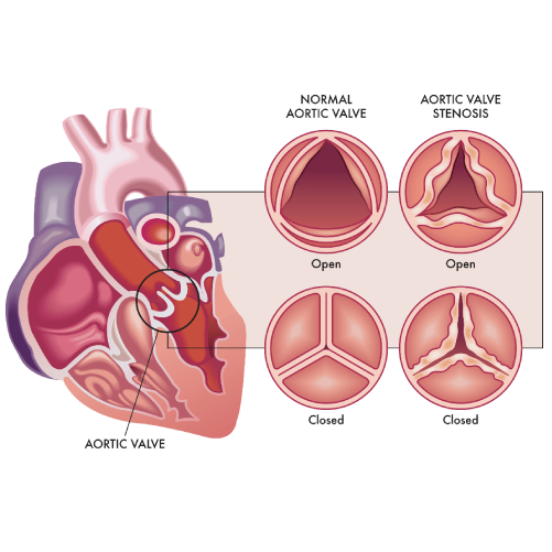
    Aortic Stenosis

    Coronary angioplasty

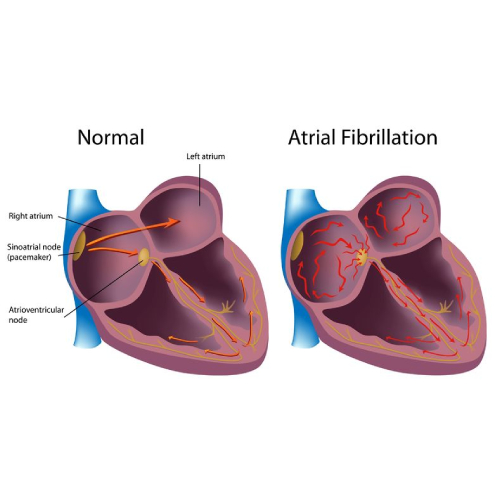
    Atrial Fibrillation

    Coronary angioplasty

    Atrial Septal Defect


    View More Services


    disease-condtions-mitral_stenosis.jpg

    Mitral stenosis is a form of valvular heart disease caused by the narrowing of the mitral valve. Mitral valve lies between the left atrium and left ventricle of the heart which is made up of two flaps of tissue called leaflets.


    cci-coronary_angioplasty.jpg


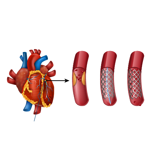
    Coronary arteries are the blood vessels that delivers oxygenated blood to the heart muscle. But, sometimes these blood vessels may get narrowed or blocked due to accumulation of cholesterol plaque. To open the blockage and restore the function of arteries, coronary angioplasty is performed.

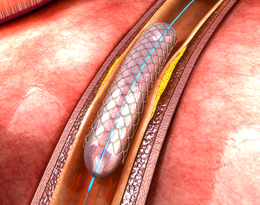
    A coronary angioplasty involves the temporary insertion of a tiny balloon inside the artery to open the blockage. It is usually combined with the placement of a stent (a metal mesh tube) to widen the artery and prevent the further chance of narrowing.

    coronary angioplasty

    Why is coronary angioplasty done ?

    Coronary angioplasty is done to treat narrowing or blockage of the heart’s blood vessels:

    • during or after a heart attack.
    • Blood vessel narrowing leading to poor heart function.
    • For relief of chest pain (angina) due to reduced blood supply to heart.

    Risks of coronary angioplasty

    Usually, it is a safe procedure, but like all other procedures, it also carries certain risks like:

    Common Issues/Risks after angiography
    Bruising Common to have bruise at groin or arm Lasts for few weeks Not to be worried about
    Allergic reactions Usually related to contrast medicine used History of previous allergy – higher risk Present as rash and headache
    Bleeding at the site of entry (Hematoma) Usually subsides in a few days But in case swelling is increasing and painful contact your physician immediately


    Serious complications after coronary angiography Less than 1 in 1000 chance People with severe heart disease are at high risk Discuss with Cardiologist before procedure
    Heart attack A serious medical emergency where the heart’s blood supply is suddenly blocked
    Stroke A serious medical condition that occurs when the blood supply to the brain is interrupted
    Loss of blood supply to limb Damage to the artery in the arm or groin in which the catheter was inserted, with the blood supply to the limb possibly being affected
    Kidney injury Damage to the kidneys caused by the contrast dye
    Radiation injury Tissue damage caused by X-ray radiation if the procedure is prolonged

    What happens before the procedure?

    A physical examination and blood test are done to evaluate the overall health condition. The person needs to follow instructions provided before undergoing the procedure:

    1. Inform the doctor about:

    • Any allergies
    • The use of current medicines, including vitamin and mineral supplements
    • Any blood disorder
    • Any surgery you may had
    • The past and present medical condition

    2. Medicines:

    Tell the doctor if you are using blood thinners or any anti-diabetic medicines. The doctor will either advise you to stop or change the dose of the medication.

    3. Food and fluid restrictions:

    • Avoid heavy meals, such as meat, fried or fatty foods eight hours before the procedure. Take light foods such as toast and cereal.
    • Fast for six hours before the procedure.
    • Stay hydrated, drink plenty of fluid up to two hours before the procedure.

    In most of the cases, you will be discharged on the same day of the hospital, so ask someone to accompany you to the hospital.

    What happens during the procedure?

    Local anaesthesia would be administered to numb the area where the catheter would be inserted. Mostly, a catheter is inserted through the wrist, and sometimes through the groin. Under fluoroscopy (a type of X-ray), the catheter will be guided to the diseased artery. Then a special medicine (called contrast or dye) will be injected into the blood vessels supplying the heart. This will delineate the presence of obstruction to blood flow. 
    Then a wire is passed across the obstruction and a balloon is inflated at the site of obstruction to compress the obstructing plaque in the artery. The balloon expansion will compress the plaques into the wall of the blood vessel and improve the blood flow. Once the blood flow is improved, the catheter will be removed, and a stent would be placed to prevent the risk of further blockage. Finally, the catheter will be removed and the puncture site is sealed.

    What to expect after the procedure?

    The nurse will check the vitals of the patient. Depending on the condition, the doctor would advise you the hospital stay. You will be advised not to bend or cross the leg for a few days if the procedure is performed through the groin region. Additionally, some other tests like X-rays and an electrocardiogram (ECG) would be done to check the condition. Before discharge, the doctor will give you certain instructions regarding wound care, medications and lifestyle.

    Recovery after coronary angioplasty

    Recover quickly and keep your heart healthy by following these tips:

    • Quit smoking.
    • Limit the intake of alcohol.
    • Take the prescribed medicines.
    • Check the cholesterol levels regularly.
    • Maintain a healthy body weight.
    • Have a healthy and well-balanced diet.
    • Exercise regularly.

    Know your numbers

    It’s important to periodically monitor and maintain the following numbers to prevent future cardiac events

    • LDL cholesterol – less than 55 mg/dL
    • Blood pressure – less than 120/80 mm Hg
    • Blood sugar – HbA1C less than 7.0%

    Book Online Consultaion


      Download: Coronary Angioplasty Blog

      GET DOWNLOAD PDF NOW

      Subscribe the Hearty Life Blogs


        Dr raghu logo


        Dr raghu logo


        Call us now if you are in a medical emergency need, we will reply swiftly and provide you with a medical aid.


        For Appointment with Dr Raghu: +91 80085 36699
        Dr Raghu’s Patient Coordinator: +91 72860 10203
        Dr Raghu’s Office Experience: +91 90001 65962
        Dr Raghu’s Social Media Communication Channel: +91 97046 51708

        Dr. Raghu | Heart Specialist in Hyderabad
        Yashoda Hospitals, Raj Bhavan Road, Somajiguda, 1st floor, Room No. 115, Hyderabad -500082, Telangana

        Best Cardiologists in Hyderabad


        © 2024, Dr. Raghu. All rights reserved. Design & Developed by AMSDigital.in

        [gtranslate]
        +91-9542475650