hand-holding-blood-glucose-meter-measuring-blood-sugar-background-is-stethoscope-chart-file-1-1.jpg

Diabetes mellitus is a chronic condition that affects more than 37 million Americans. By 2030, the disease will likely be prevalent in 1 in 10 people worldwide. Often known as a silent killer, diabetes can lead to life-threatening health complications if left untreated.

Prolonged diabetes can damage various organs of the body, including the eyes, kidneys, and skin. Also, it can lead to nerve damage (a condition known as diabetic neuropathy). However, one of the most catastrophic complications of diabetes is an increased risk of heart disease.

In this article, we’ll take a closer look at the link between diabetes mellitus and heart disease. Also, we’ll discuss how people with diabetes can lower their risk of developing cardiovascular diseases. Let’s get started.

Diabetes Mellitus and Heart Disease: Understanding the Connection


When an individual has diabetes, their body doesn’t metabolize glucose properly, which leads to elevated blood glucose levels. The excess sugar in the blood can stick to the walls of blood vessels, thus damaging and narrowing them. Also, diabetes causes damage to the nerves that control the cardiovascular system.

That, in turn, requires the heart to work harder to pump blood throughout the body. Ultimately, it can result in chronic conditions, such as coronary artery disease and heart failure. Diabetes can also increase the likelihood of blood clots, thus leading to heart attacks and strokes.

It’s also worth noting that most people with diabetes also have other underlying conditions, such as hypertension and high cholesterol levels. That, in turn, increases the risk of cardiovascular diseases.

On the other hand, a history of heart disease also increases your predisposition to diabetes mellitus. That’s because most people with cardiovascular ailments have pre-existing conditions, such as high blood pressure and obesity, which are linked to increased diabetes risk.

Minimizing the Risk of Heart Disease

Once you realize the close link between diabetes and cardiology, it’s easy to feel overwhelmed. The good news is that many of the techniques to manage and treat diabetes also help keep heart disease at bay.

Here are a few tips to help you get started:

Control Blood Sugar Levels

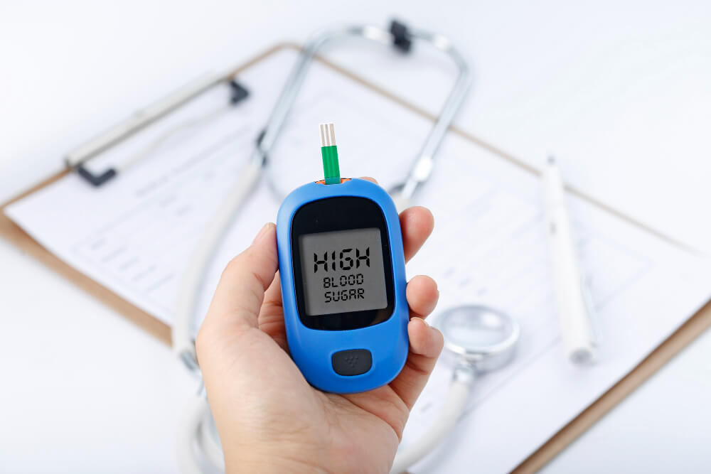
It’s crucial to undergo regular HbA1c and fasting blood glucose tests to monitor your blood sugar levels. You can even use a glucometer to test your blood glucose levels at home. Also, consult a diabetes mellitus specialist to understand whether you need to take any medications or insulin injections to keep your blood sugar levels in check.

Maintain a Healthy Body Weight

Being overweight or obese is a risk factor for both diabetes mellitus and heart disease. That makes it crucial to control your body weight. If you have a higher body mass index (BMI), consult a fitness expert or nutritionist to create a holistic weight loss plan.

Embrace a Balanced Diet

A balanced, nutrient-rich diet will help maintain your blood sugar levels and facilitate weight loss. Also, it can help keep your blood pressure and cholesterol levels in check. Avoid sugar-rich packaged and processed foods. Instead, include more fruits, vegetables, and whole grains in your diet. It’s a good idea to consult a nutritionist for a DASH or Mediterranean diet plan.

Get Regular Exercise

Exercise offers several health benefits, including weight control and emotional well-being. You can work out at the gym, go for occasional hikes, or try doing yoga. The key is to incorporate some kind of physical activity into your daily routine.

Besides these measures, you should also get your blood pressure and cholesterol regularly checked. Talk to your doctor to understand whether you need to take any medications, such as beta-blocker or statins, to lower your risk of diabetes complications further.

Safe diabetic medicines for heart disease individuals 

Conclusion

There is a clear link between diabetes mellitus and heart disease. People with diabetes are at an increased risk of developing heart disease due to the effects of high blood sugar on the blood vessels and heart. Proper management of diabetes and heart disease risk factors is essential to prevent the development of heart disease in people with diabetes.

Dr. C Raghu is an eminent cardiologist based in Hyderabad. If you or anyone you know has been diagnosed with diabetes and is at risk of developing heart disease, feel free to book a consultation with Dr. Raghu today.

Book Online Consultaion


    Diabetes Mellitus and Heart Disease Blog

    DOWNLOAD PDF

    Subscribe the Hearty Life Blogs


      Diastolic-heart-failure-img.jpg

      October 1, 2022 AbhijitHeart Failure

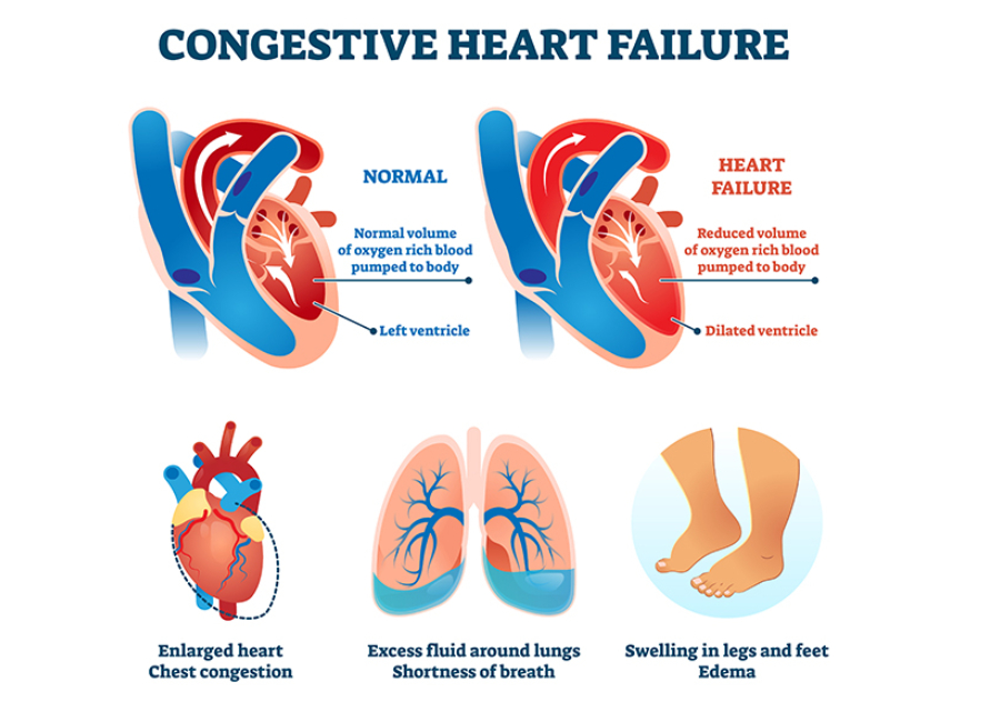
      In our previous articles, we’ve discussed the symptoms of heart failure and the steps to diagnose the same. However, the course of treatment varies for every patient based on the type of heart failure they’ve developed.

      Depending on the part of the heart’s pumping cycle that’s been affected, there are two types of heart failure. In this blog, we’ll take a closer look at diastolic dysfunction and its symptoms.

      What Causes Diastolic Dysfunction?

      diastolic dysfunction

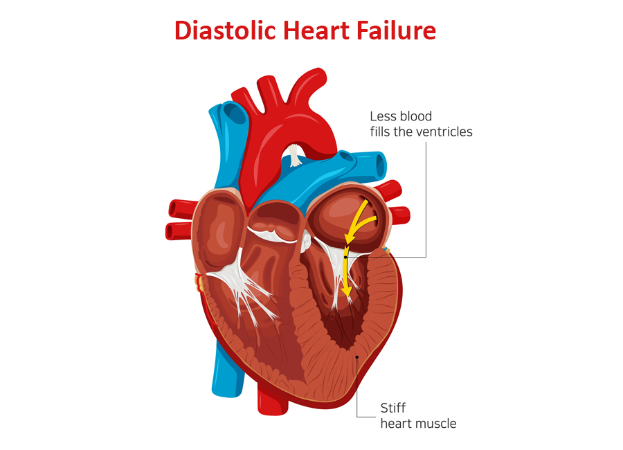
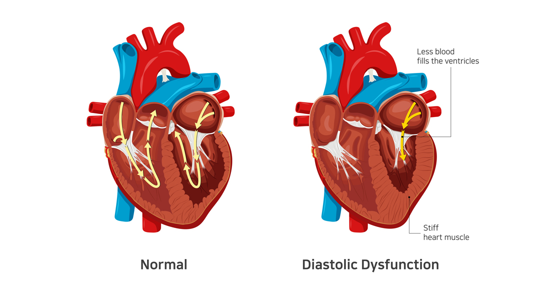
      The diastolic phase refers to the part of the heart’s pumping cycle when the ventricles (lower chambers) relax and let blood flow in from the atria (upper chambers). Diastolic dysfunction is a condition in which the ventricles don’t relax enough. That, in turn, prevents the normal amount of blood from entering the heart.

      Diastolic dysfunction is caused when the heart muscles become thicker and stiffer than usual. It’s more common in older women with hypertension and diabetes. If left untreated, it can lead to diastolic heart failure (also known as heart failure with preserved ejection fraction).

      Related : Understanding Congestive Heart Failure Symptoms

      What Does Preserved Ejection Fraction Mean?

      Ejection fraction refers to the volume of blood pumped out from the heart’s left ventricle with each contraction. For a healthy heart, the number falls in the range of 55% to 65%. A lower ejection fraction is one of the most common indicators of heart failure.

      However, it’s worth noting that many people with diastolic dysfunction have an ejection fraction of 50% or more (which is known as preserved ejection fraction). That means the left ventricle expels an adequate amount of oxygenated blood.

      However, the heart muscle doesn’t relax enough to let a sufficient quantity of blood in. That, in turn, causes the excess blood to back up in the lungs and results in fluid buildup in the feet and abdomen.

      How to differentiate systolic from diastolic dysfunction ?

      diastolic dysfunction

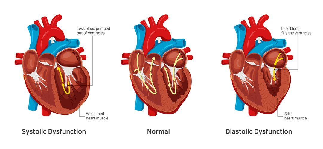
      In contrast to systolic dysfunction where the heart muscle is “weak”, in diastolic dysfunction the heart is “stiff”. This means that the heart is unable to pump blood out of the heart in systolic dysfunction whereas the heart is unable to accept further blood in diastolic dysfunction. Both conditions lead to congestion or fluid accumulation in various organs of the body. Differentiation of heart failure from systolic and diastolic dysfunction is not possible as both diseases present with similar symptoms. 

      Which conditions lead to Diastolic dysfunction?

      • Diastolic dysfunction appears consequent to uncontrolled or long-standing diabetes
      • Hypertension
      • Obesity as well as elderly people
      • Women and atrial fibrillation

      The best way to prevent and treat diastolic dysfunction is by effective control of the diseases mentioned above.

      Symptoms of Diastolic Dysfunction

      The most common symptom of diastolic dysfunction is congestion and shortness of breath due to the buildup of blood and fluid in the lungs. Breathing difficulties can get particularly worse during exertion or when lying.

      Other symptoms of diastolic dysfunction include:

      • Coughing and wheezing (due to lung congestion)
      • Loss of appetite and nausea (due to fluid buildup around the liver and in the stomach)
      • Swollen feet, legs, and abdomen (due to fluid accumulation)

      If you experience any of the given symptoms, it’s crucial to consult a doctor for a proper diagnosis.

      Treatment of Diastolic Dysfunction

      Treatment of diastolic dysfunction involves a combination of medications (diuretics or water pills) and lifestyle changes. In severe cases, a patient might need left ventricular assist devices or a heart transplant.

      Is Diastolic Dysfunction Serious?

      In the long run, diastolic dysfunction can lead to diastolic heart failure. That, in turn, increases your risk of hospitalization and death. Therefore, you should pay close attention to your symptoms and reach out to a doctor whenever you notice anything unusual.

      Dr. C Raghu is a renowned cardiologist who specializes in interventional cardiology. He has decades of experience in treating patients with different heart conditions. If you or anyone you know has developed symptoms like shortness of breath, swollen feet, loss of appetite, etc., contact Dr. Raghu to explore your treatment options.

      Book Online Consultaion


        What Are the Symptoms of Diastolic Dysfunction ? – Blog

        DOWNLOAD PDF

        Subscribe the Hearty Life Blogs

          dr. raghu

          DR. RAGHU | Best Cardiologist in Hyderabad

          MD, DM, FESC, FACC, FSCAI

          Cardiology Coronary, Vascular and

          Structural Interventions


          Conditions & Diseases

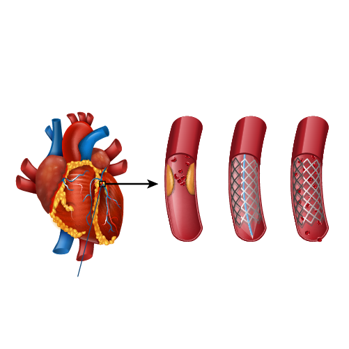
          Coronary angioplasty

          Angioplasty

          Coronary angioplasty

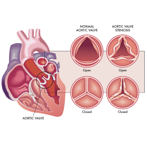
          Aortic Stenosis

          Coronary angioplasty

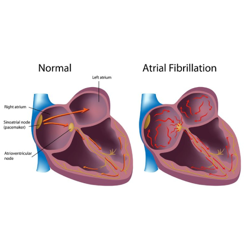
          Atrial Fibrillation

          Coronary angioplasty

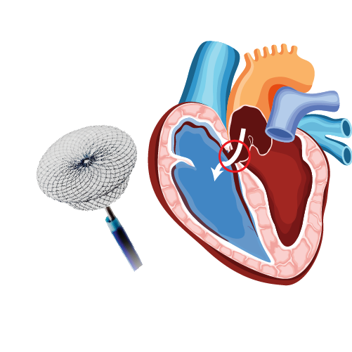
          Atrial Septal Defect


          View More Services


          Cong.-Heart-Disease.jpg

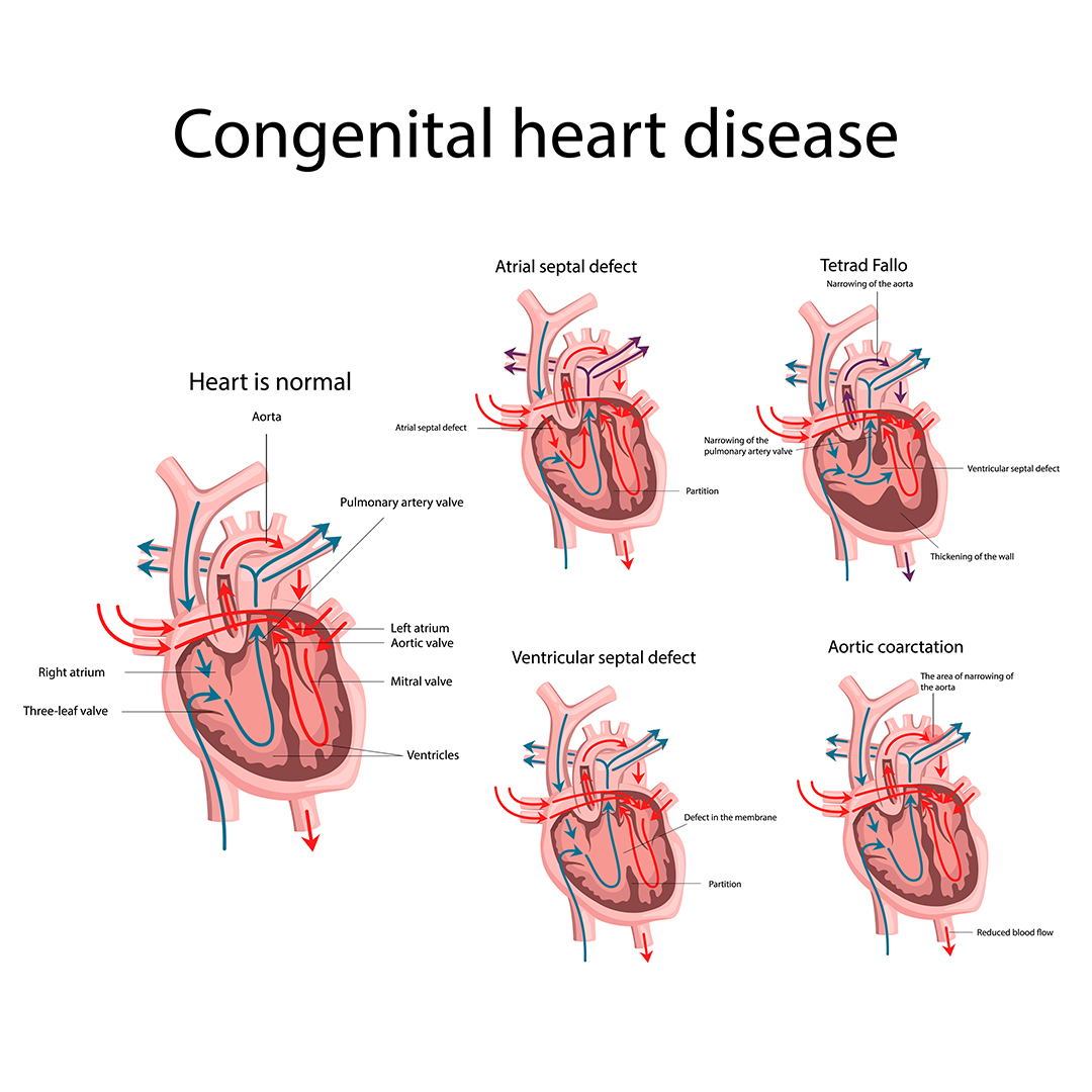
          Congenital heart diseases are diseases that are present from birth and affect the normal heart functioning. These are the most frequently occurring congenital disorder, responsible for 28% of all congenital birth defects. The birth prevalence of CHD is reported to be 8-12/1000 live births. Considering a rate of 9/1000, about 1.35 million babies are born with CHD each year globally.

          What are the Causes of Congenital Heart Defects?

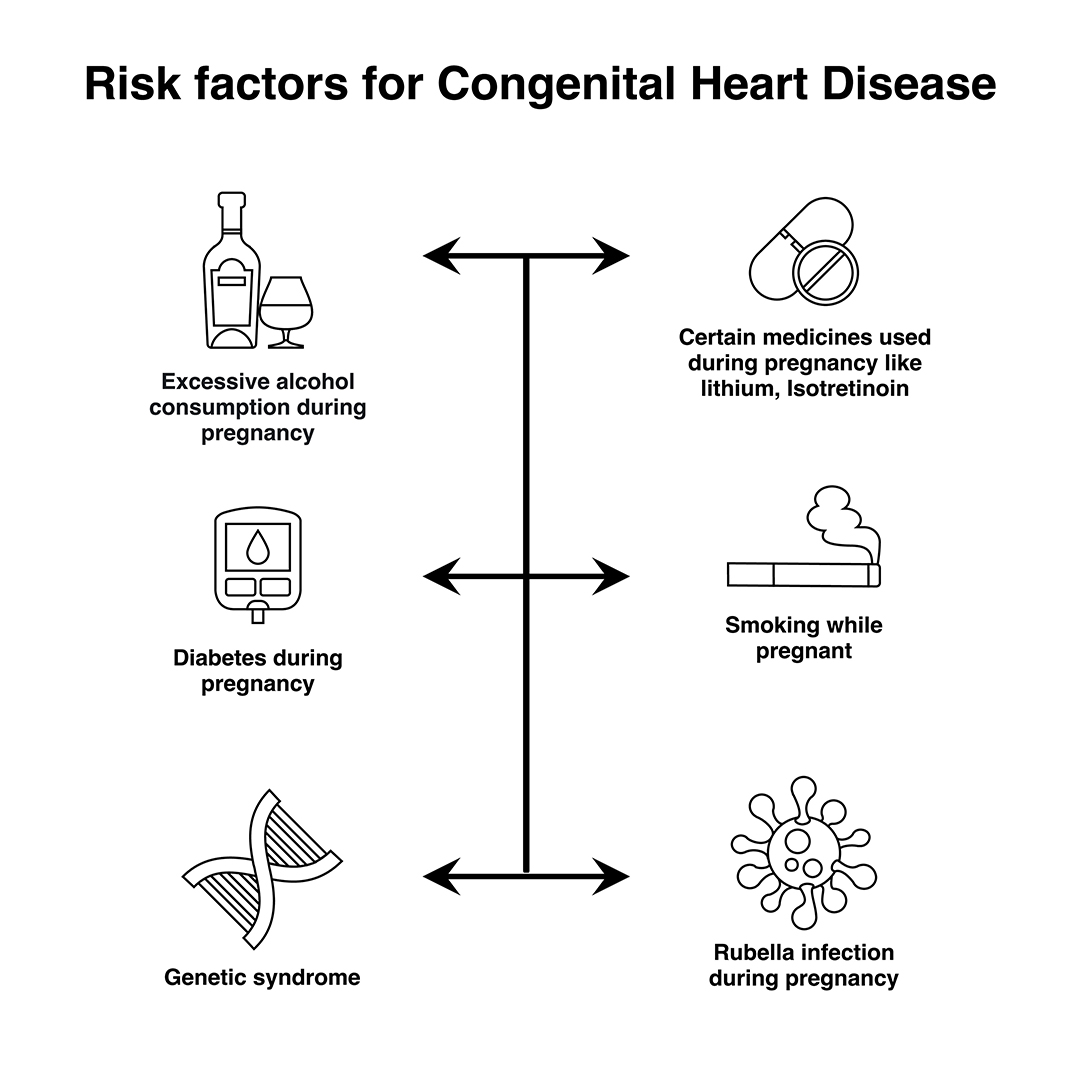
          In most cases, no obvious cause of congenital heart disease is identified. However, some things are known to increase the risk of the condition, including:

          • Down’s syndrome – a genetic disorder that affects a baby’s normal physical development and causes learning difficulties
          • the mother having certain infections, such as rubella, during pregnancy
          • the mother taking certain types of medicine during pregnancy, including statins and some acne medicines
          • the mother smoking or drinking alcohol during pregnancy
          • the mother having poorly controlled type 1 diabetes or type 2 diabetes
          • other chromosome defects, where genes may be altered from normal and can be inherited (run in the family)

          Many cases of congenital heart disease can be diagnosed before a baby is born during an ultrasound scan in pregnancy. At times a focused foetal heart scan called foetal echocardiography in specialized centers helps to diagnose the cardiac problem before birth. However, it’s not always possible to detect congenital heart defects in this way.

          congenital heart disease

          Signs and Symptoms of Congenital Heart Disease

          Congenital heart disease can have a number of symptoms, particularly in babies and children, including:

          • Rapid heartbeat
          • Rapid breathing
          • Swelling of the legs, tummy or around the eyes
          • Extreme tiredness and fatigue
          • A blue tinge to the skin or lips (cyanosis)
          • Tiredness and rapid breathing when a baby is feeding

          These problems are sometimes noticeable soon after birth, although mild defects may not cause any problems until later in life.

          Types of Congenital Heart Disease

          There are many types of congenital heart disease and they sometimes occur in combination. Some of the more common defects include:

          • Septal defects – where there’s a hole between 2 of the heart’s chambers (commonly referred to as a “hole in the heart”)
          • Coarctation of the aorta – where the main large artery of the body, called the aorta, is narrower than normal
          • Pulmonary valve stenosis – where the pulmonary valve, which controls the flow of blood out of the lower right chamber of the heart to the lungs, is narrower than normal
          • Transposition of the great arteries – where the pulmonary and aortic valves and the arteries they’re connected to have swapped positions
          • Underdeveloped heart – where part of the heart doesn’t develop properly making it difficult for it to pump enough blood around the body or lungs.

          congenital heart disease

          Treating Congenital Heart Disease

          Treatment for congenital heart disease usually depends on the defect you or your child has.

          Mild defects, such as holes in the heart, often don’t need to be treated, as they may improve on their own and may not cause any further problems.

          Surgery or interventional procedures are usually required if the defect is significant and causing problems. Modern surgical techniques can often restore most or all of the heart’s normal function.

          However, people with congenital heart disease often need treatment throughout their life and therefore require specialist review during childhood and adulthood. This is because people with complex heart problems can develop further problems with their heart rhythm or valves over time.

          Most surgery and interventional procedures aren’t considered to be a cure. The affected person’s ability to exercise may be limited and they may need to take extra steps to protect themselves from getting infections.

          It’s important that a person with heart disease and their parents or carers discuss these issues with their specialist medical team.

          Book Online Consultaion


            Basics of Congenital Heart Disease Blog

            DOWNLOAD PDF

            Subscribe the Hearty Life Blogs

              dr. raghu

              DR. RAGHU | Best Cardiologist in Hyderabad

              MD, DM, FESC, FACC, FSCAI

              Cardiology Coronary, Vascular and

              Structural Interventions


              Conditions & Diseases

              Coronary angioplasty

              Angioplasty

              Coronary angioplasty

              Aortic Stenosis

              Coronary angioplasty

              Atrial Fibrillation

              Coronary angioplasty

              Atrial Septal Defect


              View More Services


              disease-condtions-device_closure_for_asd.jpg

              January 16, 2021 Dr. RaghuHearty Life


              An atrial septum is a muscular wall that separates the upper chambers of the heart called atria. An ASD is a common congenital heart disease where the septum is not formed properly producing a left-to-right shunt, which leads to mixing of oxygenated and deoxygenated blood. This causes pulmonary hypertension and right heart enlargement.

              What are the treatment options for ASD?

              Small atrial defects do not need any treatment and close on its own. Even in adulthood small ASDs may remain asymptomatic. Some large defects that persists in adulthood may become symptomatic and need closure. The ASDs can be closed by:

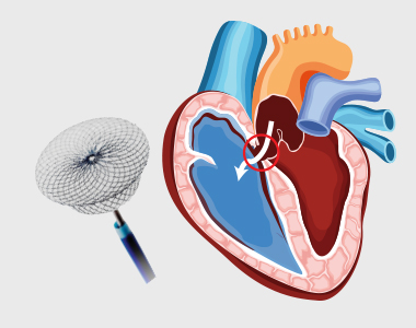
              • Percutaneous closure using a device
              • Closure through open heart surgery

              Percutaneous device closure is the preferred treatment for certain defects type.

              What is Percutaneous Closure of Atrial Septal Defect (ASD)?

              A noninvasive procedure known as percutaneous transcatheter approach is considered depending on the size and severity of the defect. Moderate to large-sized ASD along with pulmonary hypertension requires to be closed. The procedure is performed by inserting a special closure device either folded or attached to a catheter into the vein of the leg and is advanced to the heart through the defect, which closes the hole by a special mechanism.

              Are there risks associated with the procedure?

              The success rate of the procedure is about 95%. But the risks involved, and their estimated incidence of occurrence include:

              • Device dislodgement leading to emergency heart surgery: 1%
              • Device erosion (device eroding through the heart walls): 0.3%
              • Death: less than 1% usually due to perforation of the heart chamber
              • Dislodgement of clot or air bubbles to the brain leading to other organs: less than 1%
              • Arrhythmia: 1 to 2%
              • Allergic dye reaction
              • Anaesthetic reaction
              • Injury to the artery/vein/nerves in the groin
              • Perforation of the oesophagus
              • Infection
              • Allergic reaction to the nickel component of the device

              Patients with small ASD may not develop any complications, but large-sized defects may lead to serious complications which demands surgery and prolonged hospitalization.

              What is the pre-procedure work-up?

              Pre-operative tests to assess the general health of the patient include:

              • Chest x-ray
              • Electrocardiogram
              • Blood tests
              • Kidney function

              A detailed diagnosis of the defect should be performed which includes transthoracic and transoesophageal echocardiogram used to assess the size, location and the suitability of the procedure.

              How should I prepare for the procedure?

              • Patients wearing dentures, glasses or a hearing assist device can plan to wear them during the procedure.
              • Patient will be instructed about dietary restriction to be followed before the procedure.

              Are there any specific instructions about medications?

              • The healthcare provider may ask you to stop certain medications, such as warfarin or other blood thinners.
              • If diabetic, consult the physician about how the medication needs to be adjusted.
              • Provide information about specific allergies regarding iodine, shellfish, X-ray dye, latex or rubber products etc.

              What happens during the procedure?

              • The patient might be asked to have a shower before the procedure.
              • The patient is asked to wear a hospital gown and lie on an X-ray table where an X-ray camera will move over the chest during the procedure.
              • Arrangements for intravenous administration of medications or fluids during the procedure will be made.
              • The site where the catheter will be inserted is cleaned and sterile drapes were used to cover the site to prevent the infection.
              • Electrodes will be placed on the chest and are attached to an electrocardiograph monitor (ECG).
              • A sedative might be given to relax, and a local anesthetic is given to numb the site of catheter introduction.
              • A plastic sheath will be inserted in the groin, through which a catheter is inserted and threaded to the heart.
              • The physician may also inject a dye which may make you feel hot or flushed for several seconds. Inform the doctor if there is an allergic reaction like itching or tightness in throat, nausea and chest discomfort.
              • The X-ray cameras are used to obtain the measurements of pressures and oxygen content in the chambers.
              • The appropriate size and the location of the closure might be visualized using a small catheter connected with an ultrasound transducer.
              • A special catheter is used to advance the device into the heart and through the defect.
              • The device is slowly pushed out of the catheter allowing each side of the device to open and close each side of the hole in the septum.
              • The proper position of the device is ensured and is released from the catheter.
              • It may take 1-2 hours for the procedure, but preparations must be made to spend about 5-9 hours in the hospital.
              • The patient should be accompanied by someone who can drive him home, as the patient will not be allowed to drive on the same day.

              What care should be taken after the procedure?

              • The catheter and the imaging probe are removed after the completion of the procedure.
              • Pressure on the incision site or occasionally a small suture is used to close the vein.
              • Bed rest is advised for several hours to prevent bleeding, but call the doctor if you notice any bleeding.
              • You might be advised to drink plenty of water to wash out the contrast material from the body.
              • Your heart rate and rhythm are monitored; you may be asked to stay overnight in the hospital.
              • Medications, such as aspirin are prescribed to prevent blood clots.
              • Strenuous activity and heavy lifting should be avoided for at least six months.
              • Antibiotic prophylaxis is required for at least six months or lifelong to prevent endocarditis, as per doctor’s advice.


              • 24 hours emergency services
              24 hours emergency services
              • Clinics- weekly basis/monthly basis/ Yearly basis
              Clinics- weekly basis/monthly basis/ Yearly basis
              • Prevention of cardiovascular diseases
              Prevention of cardiovascular diseases
              • Diagnosis
              Diagnosis

              BOOK AN APPOINTMENT

                dr. raghu

                Dr. RAGHU

                MD, DM, FESC, FACC, FSCAI

                Cardiology Coronary, Vascular and

                Structural Interventions


                Conditions & Diseases

                Coronary angioplasty

                Angioplasty

                Coronary angioplasty

                Aortic Stenosis

                Coronary angioplasty

                Atrial Fibrillation

                Coronary angioplasty

                Atrial Septal Defect


                View More Services


                Dr raghu logo


                Dr raghu logo


                Call us now if you are in a medical emergency need, we will reply swiftly and provide you with a medical aid.


                For Appointment with Dr Raghu: +91 80085 36699
                Dr Raghu’s Patient Coordinator: +91 72860 10203
                Dr Raghu’s Office Experience: +91 90001 65962
                Dr Raghu’s Social Media Communication Channel: +91 97046 51708

                Dr. Raghu | Heart Specialist in Hyderabad
                Yashoda Hospitals, Raj Bhavan Road, Somajiguda, 1st floor, Room No. 115, Hyderabad -500082, Telangana

                Best Cardiologists in Hyderabad


                © 2024, Dr. Raghu. All rights reserved. Design & Developed by AMSDigital.in

                [gtranslate]
                +91-9542475650